ILIOSACRAL SCREW FIXATION FOR POSTERIOR PELVIC RING INSTABILITY: SURGICAL TECHNIQUE, IMAGING PROTOCOLS, AND COMPLICATION MANAGEMENT
- K. C. MATHEW , Professor and Head of the department, DEPARTMENT OF ORTHOPAEDICS, SREE MOOKAMBIKA INSTITUTE OF MEDICAL SCIENCES, KULASEKHARAM, KANYAKUMARI -629161, TAMILNADU, INDIA.
- ZAKIR HUSSAIN MOHAMED , Postgraduate Resident, DEPARTMENT OF ORTHOPAEDICS, SREE MOOKAMBIKA INSTITUTE OF MEDICAL SCIENCES, KULASEKHARAM, KANYAKUMARI -629161, TAMILNADU, INDIA.
- ANTO JEFRIN VISHAL A , Senior Resident, DEPARTMENT OF ORTHOPAEDICS, SREE MOOKAMBIKA INSTITUTE OF MEDICAL SCIENCES, KULASEKHARAM, KANYAKUMARI -629161, TAMILNADU, INDIA.
- ANNAMALAI T T , Postgraduate Resident, DEPARTMENT OF ORTHOPAEDICS, SREE MOOKAMBIKA INSTITUTE OF MEDICAL SCIENCES, KULASEKHARAM, KANYAKUMARI -629161, TAMILNADU, INDIA.
- ROHIN G , Postgraduate Resident, DEPARTMENT OF ORTHOPAEDICS, SREE MOOKAMBIKA INSTITUTE OF MEDICAL SCIENCES, KULASEKHARAM, KANYAKUMARI -629161, TAMILNADU, INDIA.
- GOWTHAM RAJ G , Postgraduate Resident, DEPARTMENT OF ORTHOPAEDICS, SREE MOOKAMBIKA INSTITUTE OF MEDICAL SCIENCES, KULASEKHARAM, KANYAKUMARI -629161, TAMILNADU, INDIA.
- KAVIN RAJA S , Postgraduate Resident, DEPARTMENT OF ORTHOPAEDICS, SREE MOOKAMBIKA INSTITUTE OF MEDICAL SCIENCES, KULASEKHARAM, KANYAKUMARI -629161, TAMILNADU, INDIA.
Article Information:
Abstract:
Background: Pelvic ring injuries represent a significant proportion of skeletal trauma, with nearly 40% classified as unstable due to posterior ring disruption. Sacroiliac (SI) joint injuries carry substantial morbidity, including chronic pain, neurologic dysfunction, and impaired quality of life. While open reduction and internal fixation (ORIF) was historically standard, the introduction of percutaneous iliosacral screw fixation has offered a less invasive alternative with comparable biomechanical stability. Methods: This review synthesizes the available surgical and anatomic literature on percutaneous iliosacral screw fixation, focusing on indications, preoperative planning, intraoperative technique, fluoroscopic imaging protocols, complication profiles, and postoperative outcomes.Results and Discussion: Iliosacral screws are safe and effective across a broad range of sacral morphologies, including dysmorphic variants, when appropriate preoperative planning and fluoroscopic technique are employed. Key complications include neurovascular injury, malreduction, and malunion. Screw malposition rates may approach 24% and neurologic sequelae up to 18% have been reported, underscoring the importance of meticulous technique.Conclusion: Iliosacral screw fixation remains a versatile, minimally invasive technique for posterior pelvic ring stabilization. Understanding sacral anatomy, recognizing dysmorphic variants, and obtaining optimal intraoperative imaging are critical determinants of success.
Keywords:
Article :
INTRODUCTION:
Pelvic ring injuries, while accounting for approximately 3% of all skeletal fractures, carry disproportionate clinical consequences due to the intimate proximity of the posterior pelvis to critical neurovascular structures, viscera, and the lumbosacral plexus.1 Approximately 40% of these injuries are classified as mechanically unstable owing to posterior ring disruption.2 Injury to the sacroiliac (SI) joint, in particular, is associated with a constellation of debilitating sequelae, including chronic pelvic pain, sexual dysfunction, bowel and bladder impairment, and significant impairment in the ability to return to occupational activity. 3–7
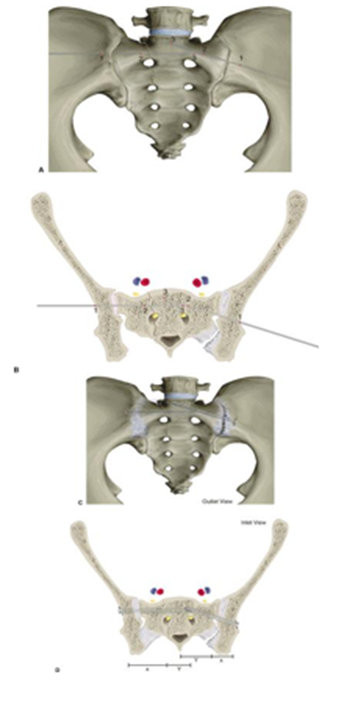
Figure 1. Anatomical schematic demonstrating iliosacral screw fixation pathways and safe osseous corridors within the posterior pelvic ring.
Historically, the definitive surgical management of unstable posterior pelvic ring injuries was accomplished via open reduction and internal fixation (ORIF) utilizing sacral bars or posterior plating constructs. Although effective biomechanically, open procedures carry substantial risks,8-10, including extensive soft tissue dissection, prominent implant profiles, iatrogenic nerve injury, infection, and considerable intraoperative blood loss, compounding the physiologic insult in an already traumatized patient.6,11–13
The development of percutaneous fixation via iliosacral screws has substantially altered the management paradigm for these injuries. Compared with open techniques, percutaneous iliosacral screw insertion is associated with reduced operative time, diminished soft tissue disruption, and lower intraoperative blood loss.14 Additionally, iliosacral screws demonstrate considerable versatility: they can be employed in multiple patient positions (supine, prone, lateral), utilized across a spectrum of sacral fracture morphologies and SI joint disruptions, and safely deployed even in patients with sacral dysmorphism when appropriate precautions are taken.15–19
Despite these advantages, the technique demands a thorough understanding of sacropelvic anatomy, fluoroscopic imaging principles, and fracture reduction strategies. The rate of screw malposition may approach 25%, and neurologic injury rates as high as 18% have been documented in the literature.6,20–25 This review comprehensively addresses the indications, surgical technique, imaging protocols, complications, and expected outcomes associated with percutaneous iliosacral screw fixation of the posterior pelvic ring.
Indications and Contraindications
Indications
Iliosacral screws may be employed as standalone fixation or as supplemental stabilization in conjunction with anterior pelvic fixation.26,27 Originally described for isolated SI joint dislocations and fracture-dislocations,28 their applications have expanded to encompass:
· Unstable posterior pelvic ring injuries
· Sacroiliac joint dislocation and fracture-dislocation
· Spinopelvic dissociation
· Incomplete sacral fractures (Denis zones 1–3) with or without pelvic instability
· Vertical shear posterior pelvic fractures
· Sacral fractures demonstrating persistent gapping following anterior symphyseal plating
The minimally invasive nature of iliosacral screw insertion renders it particularly advantageous in the context of extensive soft tissue trauma, open fractures, and degloving injuries, where conventional plating would require dissection through compromised tissue.16,29 Anterior-posterior compression (APC) injuries with bladder disruption or contaminated anterior soft tissue can be managed with anterior external fixation supplemented by posterior iliosacral screws. APC type IIb injuries — characterized by posterior SI ligament attenuation with sagittal plane instability — may be addressed with iliosacral screws combined with anterior symphyseal plating.30
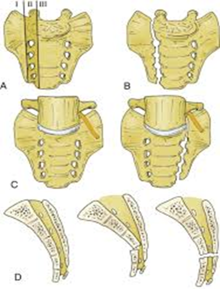
Figure 2. Schematic illustration demonstrating the Denis classification of sacral fractures. Zone I fractures involve the sacral ala lateral to the neural foramina, Zone II fractures extend through the foramina, and Zone III fractures involve the central sacral canal with the highest risk of neurologic injury
Table 1. Indications and Contraindications for Percutaneous Iliosacral Screw Insertion
|
Category |
Details |
|
Indications |
Unstable posterior pelvic ring injuries; SI joint dislocation; spinopelvic dissociation; incomplete sacral fractures ± pelvic ring instability; vertical posterior pelvic fractures; sacral fractures with gapping after symphyseal plating |
|
Contraindications |
Delayed fixation (>5 days); active surgical site infection; severe sacral dysmorphism; morbid obesity; horizontal sacral fractures |
SI = sacroiliac.
Contraindications
Absolute contraindications to closed reduction and percutaneous fixation include an inability to achieve satisfactory closed reduction and the presence of active infection at the proposed surgical site. A delay to definitive fixation exceeding five days constitutes a relative contraindication, as organized hematoma may preclude accurate fracture reduction.31 Horizontal sacral fractures are poorly suited for iliosacral screw constructs, as the implant would be oriented parallel to the fracture line, providing insufficient fixation.
Historically, transitional lumbosacral anatomical variants were regarded as relative contraindications16; however, subsequent investigations have confirmed that the majority of patients with sacral anomalies can safely undergo percutaneous fixation with appropriate recognition and planning.18,19 Severe sacral dysmorphism may nonetheless preclude safe screw placement. U-shaped sacral fractures with kyphotic deformity or foraminal narrowing may require a supplemental posterior open procedure for sacral reduction and nerve root decompression.17 Morbidly obese patients may present technical challenges due to suboptimal fluoroscopic visualization.
Preoperative Planning
Thorough preoperative assessment is the foundation of safe iliosacral screw insertion. A comprehensive physical examination with careful documentation of soft tissue injury, neurologic status, and associated injuries — including cranial, thoracic, abdominal, spinal, and extremity trauma — is mandatory.
Standard radiographic evaluation includes anteroposterior (AP) pelvis, AP chest, and lateral cervical spine projections for all patients with polytrauma. Advanced imaging with computed tomography (CT), utilizing axial slices oriented perpendicular to the sacral slope,32 is strongly recommended for operative planning. CT imaging identifies sacral fractures that are missed on plain radiographs in approximately 30% of cases27 and provides critical information regarding bone quality, sacral morphology, neural foraminal anatomy, and adjacent vascular structures.32
Characterization of the injury pattern guides guidewire orientation: a posterior-to-anterior trajectory is preferred for SI joint dislocations to achieve compression across the joint, whereas a transverse orientation is more appropriate for sacral fractures.30 Comminuted sacral fractures involving the foraminal pathways are best treated with fully threaded static screws rather than lag screws to mitigate the risk of nerve root entrapment from overcompression.17
Sacral Dysmorphism
Sacral dysmorphism affects an estimated 35% to 58% of the adult population and constitutes a recognized risk factor for aberrant iliosacral screw placement.18,19,34 Dysmorphic features include anomalous cranial fusion of the sacrum to L5 (sacralization), abnormal development of transverse processes and pedicles, malformed SI joints, and irregular neural foramina.18,19,35
Identification of dysmorphism begins with plain radiography. The pelvic outlet view may reveal proximal positioning of S1 collinear with the iliac crests, rudimentary mammillary bodies (underdeveloped transverse processes), acute coronal-plane alar sloping, and enlarged irregular S1 foramina.32 The iliac cortical density (ICD) the fluoroscopic landmark representing the alar slope is displaced posteriorly and caudally in the dysmorphic sacrum, potentially misleading the surgeon regarding safe screw trajectory.18,32 Detailed characterization of aberrant anatomy, including undulating SI joint contours, is best accomplished with preoperative CT.
Importantly, both morphologically normal and dysmorphic sacra can accommodate screws of at least 75 mm in length, and the safe zones for screw insertion are equivalent in both variants.18
Surgical Technique
Patient Positioning and Preparation
The procedure is performed under general anesthesia, with administration of a first-generation cephalosporin for antimicrobial prophylaxis. Spinal precautions are maintained during patient transfer to a radiolucent operating table. A soft lumbosacral support constructed from folded towels or blankets is positioned beneath the patient to elevate the pelvis and facilitate percutaneous posterior access.
Iliosacral screw insertion may be performed with the patient in the supine, prone, or lateral decubitus position. Supine positioning is generally preferred as it offers anesthesia familiarity, simultaneous access to the anterior and posterior pelvis, compatibility with polytrauma management, and potential facilitation of closed reduction maneuvers.30,33 The prone position affords improved posterior access but limits direct visualization of SI joint reduction. The lateral position is not recommended for patients with potential spinal injuries and complicates anterior pelvic access.
When distal femoral traction is required for reduction, a pulley system attached to the operating table is employed. Neurodiagnostic monitoring should be considered for patients with transforaminal sacral fractures, preoperative neurologic deficits, or cognitive impairment.29
Closed Reduction Techniques
Achieving and confirming anatomic SI joint and sacral fracture reduction is a prerequisite for accurate screw placement. Sacral and pelvic injuries with an intact posterior tension band are typically amenable to closed reduction.33 The nature of displacement guides the reduction strategy: cephalad hemipelvis migration may be corrected with distal femoral traction, while caudal migration of the contralateral hemipelvis can be counteracted with an ipsilateral traction boot. Supplementary techniques include internal rotation of both lower extremities using circumferential thigh-and-foot taping, pelvic binders or sheets for external rotation deformities, pelvic C-clamps, and Schanz pins for multiplanar percutaneous manipulation.33 If closed reduction cannot be achieved, open reduction must be performed.
4.3 Surgical Approach and Entry Point
In the supine position, the anterior superior iliac spine (ASIS) is identified by palpation and a line is drawn from this landmark directed perpendicular to the floor. A second line, drawn in alignment with the femoral shaft, creates four quadrants at their intersection. The posterosuperior quadrant delineates the optimal starting zone for iliosacral screw placement, confirmed intraoperatively with lateral sacral fluoroscopy. A stab incision is made through this zone and a smooth Kirschner wire (K-wire) is advanced through the incision until it contacts the outer table of the ilium. The instrumentation should be directed toward the osseous corridor between the anterior sacral ala and the S1 neural foramina.
In the prone position, the entry point corresponds to the intersection of a vertical line descending from the posterior superior iliac spine and a horizontal line extending proximally in line with the greater trochanter and femur.
Fluoroscopic Imaging Protocol
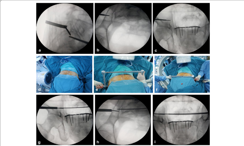
Figure 3. Intraoperative fluoroscopic imaging used during percutaneous iliosacral screw fixation. (A) Anteroposterior pelvic view confirming patient positioning and preliminary guidewire orientation. (B) Pelvic inlet view demonstrating visualization of the anterior sacral cortex and screw trajectory. (C) Pelvic outlet view delineating the sacral foraminal tunnels and safe osseous corridor for screw placement.
Intraoperative fluoroscopic imaging is central to safe screw placement. The fluoroscope is positioned on the side contralateral to the injured hemipelvis. Initial AP imaging confirms proper patient positioning and pelvic alignment. Inlet and outlet views are then customized for each patient.
The pelvic inlet view is obtained by directing the fluoroscope approximately 45° caudad, with correct positioning confirmed when the S1 vertebral body overlaps the S2 body, appearing as concentric circles. This view provides direct visualization of the anterior sacral cortex and allows detection of ventral cortical perforation by the guide pin or screw.
The pelvic outlet view is obtained by directing the beam approximately 45° cephalad, with accurate positioning confirmed when the pubic symphysis is superimposed on the S2 vertebral body. The outlet view delineates the corticated edges of the sacral foraminal tunnels and permits identification of neural foraminal perforation.
A true lateral sacral view is obtained by adjusting the fluoroscope to superimpose the greater sciatic notches bilaterally. On this view, the ICD is identified and correlated with the preoperative CT scan to confirm appropriate alar-level targeting. This image is obtainable only after accurate posterior pelvic reduction.
In patients with sacral dysmorphism, the fluoroscope is adjusted to focus on the specific sacral segment receiving the screw. Unique fluoroscopic reference points apply to each level, as the anterior borders of S1 and S2 differ in their relationships to surrounding anatomy.
Guide Pin and Screw Insertion
Following confirmation of closed reduction, a smooth or drill bit-tipped 0.62-mm K-wire is inserted under triplanar fluoroscopic control through the stab incision to the lateral iliac cortex. Threaded guide pins are avoided as they limit tactile feedback for detecting bony tunnel breaches. The starting point is secured by gently tapping the wire to engage the cortex, following which the skin incision is extended and blunt deep dissection is performed with a narrow periosteal elevator or drill guide.
The guide pin is advanced under power drill assistance through the lateral iliac cortex and across the SI articulation into the sacral ala, with continuous fluoroscopic monitoring in inlet and outlet projections. On the outlet view, the screw is directed approximately 20° cranially for normal sacra and 30° cranially for dysmorphic sacra.18 On the inlet view, the trajectory is angled approximately 5° anteriorly for normal sacra and 15° anteriorly for dysmorphic variants.18
The drill bit-tipped guide pin is preferred over a threaded pin as it provides superior tactile feedback for impending cortical perforation. For SI joint dislocations, the screw trajectory runs posterior to anterior; for sacral fractures, a transverse orientation is used.30 The guide pin is halted just cephalad to the corticated margin of the upper-sacral nerve root tunnel as identified on the outlet view. The surgeon must appreciate the three-dimensional path of the sacral nerve roots: posterior to anterior, medial to lateral, and superior to inferior.
Once the guide pin reaches the level of the sacral nerve root foramen, a true lateral sacral image is obtained by superimposing the bilateral greater sciatic notches. The pin tip must be confirmed to lie caudal to the ICD and cephalad to the interosseous course of the upper sacral nerve root.
The guide pin is then advanced to the midline of the upper sacral vertebral body. Depth measurement is performed with a reverse ruler, and a cannulated drill is advanced over the guide pin, followed by a cannulated tap when required by bone density.
A 7.0-mm cancellous partially threaded screw is selected when compression is desired (e.g., SI joint dislocation), and a fully threaded screw is used when compression is contraindicated (e.g., transforaminal sacral fractures with nerve root at risk of entrapment). Using a washer and rollover imaging confirms that the screw does not inadvertently penetrate the posterior iliac cortex, which is of particular concern in osteoporotic patients.
Frequent fluoroscopic imaging is obtained during cannulated drilling, tapping, and screw insertion to verify that the guide pin does not inadvertently advance. A 20°–30° obturator oblique view permits visualization of the tangential posterior ilium during screw tightening. Additional screws or supplementary fixation are deployed if residual instability is identified on the fluoroscopic stress examination. The percutaneous wound is irrigated and closed.
Complications and Management
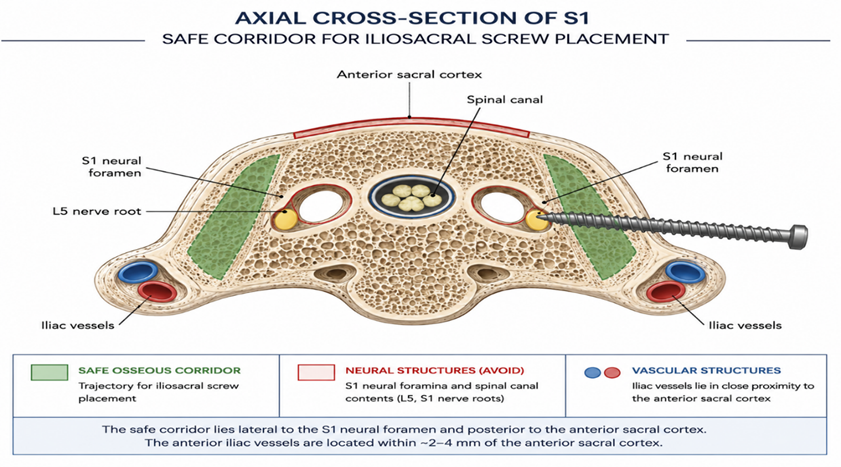
Figure 4. Axial cross-section of S1 demonstrating the safe corridor for iliosacral screw placement (green shaded zone). The trajectory must remain lateral to the S1 neural foramen and posterior to the anterior sacral cortex. The anterior iliac vessels lie within 2.4 mm of the anterior cortex
Percutaneous iliosacral screw insertion, when performed with meticulous technique and thorough preoperative planning, is a safe and effective procedure.15–17,36 However, the narrow anatomic safe zones and complex three-dimensional geometry of the sacropelvic region render the procedure susceptible to neurovascular complications with even minor deviations in screw trajectory. As little as 4° of misdirection has been shown to direct the screw into the S1 foramen or through the ventral sacral cortex.37 In cadaveric studies, the anterior cortex of the sacral ala has been found to lie within 2.4 mm of the internal iliac vein, and the L5 nerve root within 1 mm of the alar margin.38
The reported incidence of screw malposition ranges up to 24%,23,37 with neurologic sequelae occurring in up to 18% of cases, attributable to complex sacral osteology and unrecognized dysmorphism.6,23,39–41
Table 2. Complications and Management Strategies for Percutaneous Iliosacral Screw Insertion
|
Complication |
Management |
|
Iliac vessel injury |
Emergent vascular/surgical consultation; ligation, repair, retroperitoneal packing, or endovascular stenting |
|
Lumbosacral nerve injury |
Screw revision; expectant management for incomplete injuries |
|
Superior gluteal artery injury |
Angiographic embolization; ligation if accessible |
|
Postoperative malreduction |
Observation for minimal displacement; revision ORIF or SI joint fusion for significant deformity |
|
Anterior pelvic / contralateral SI instability |
Adjunctive symphyseal plating; joystick reduction with contralateral SI fixation |
|
Wound complications |
Antibiotics (superficial); incision, drainage ± implant removal (deep) |
|
Implant failure |
Observation (asymptomatic with maintained union); revision fixation (symptomatic or loss of reduction) |
|
Sacral nonunion |
Debridement, bone grafting, and revision fixation |
ORIF = open reduction and internal fixation; SI = sacroiliac.
Vascular Complications
Injury to the superior gluteal artery is a recognized complication, most commonly involving the deep superior branch, which lies in proximity to the ideal screw starting point. In a cadaveric investigation by Collinge and colleagues,42 10 of 58 percutaneously placed screws, 18%) either impaled or entrapped the superior gluteal neurovascular bundle, with a mean screw-to-bundle distance of 9.1 ± 6.8 mm. Tamponade is generally ineffective; angiographic embolization is the preferred management strategy and has been reported to be successful.20,21
The common and internal iliac veins course along the ventral surface of the sacral ala at the S1 level, lying immediately anterior to the SI joint or just medially.38 Injury to these structures mandates emergent vascular or surgical consultation; management options include ligation or primary repair, retroperitoneal packing, and endovascular stent placement.
Neurologic Complications
Neurologic injury involves the lumbosacral nerve roots and should prompt screw revision. In a study by van den Bosch and colleagues,25 7 of 88 patients (7.9%) required screw revision for neurologic injury, with complete symptom resolution in all cases. These data underscore the reversibility of neurologic deficits when screw malposition is identified and addressed promptly.
Malreduction and Malunion
Malreduction and malunion represent the most frequently encountered complications.6,14,43 In a study of vertically unstable pelvic fractures, malunion occurred in 36% of pelves treated with anterior plating combined with iliosacral screws versus 44% treated with iliosacral screws alone. While statistical significance was not achieved, the data suggest that combined fixation may reduce the risk of late displacement. Posterior pelvic ring displacement exceeding 1 cm has been definitively associated with a higher incidence of chronic pain and poorer functional outcomes.7 Revision surgery with ORIF or SI joint fusion may provide partial pain relief in symptomatic patients.6
Implant Failure, Nonunion, and Infection
Implant failure and nonunion are infrequent complications.14 When symptomatic, treatment consists of debridement, bone grafting, and revision fixation. Infection rates after percutaneous iliosacral screw insertion are remarkably low, with several studies reporting 0%,3,6,14,25 in marked contrast to the infection rates associated with open procedures.
Postoperative Care and Rehabilitation
Intravenous antibiotics are continued for 24 hours postoperatively. Chemical thromboprophylaxis is initiated on the first postoperative day and maintained for 30 days. Supervised rehabilitation is initiated by a licensed physical therapist according to a protocol individualized to the patient's overall condition and associated injuries.
The stabilized hemipelvis is protected with partial weight bearing using crutches or a walker for the initial six postoperative weeks. Progressive weight bearing follows, intending to achieve unassisted ambulation by 12 weeks. Nonimpact aerobic activity and hydrotherapy are permitted from six weeks onward.
Radiographic follow-up with inlet and outlet pelvic projections is obtained in the recovery room and at six- and twelve-week postoperative visits. A postoperative CT scan is frequently obtained to assess implant position and fracture reduction quality. Clinical follow-up appointments are scheduled at two, six, and twelve weeks, with subsequent intervals determined by patient-specific factors. Most adult patients with physically demanding occupations achieve functional return to work at four to six months postoperatively.
Clinical Outcomes
Systematic assessment of outcomes following fixation of unstable pelvic injuries is confounded by small cohort sizes, heterogeneous injury patterns and fixation strategies, variable follow-up durations, lack of standardized outcome measures, and the confounding effects of polytrauma.3,6,27,29
The neurologic outcome following sacral fracture fixation is largely determined by the Denis zone of injury: zone 3 fractures carry the highest association with bowel, bladder, and sexual dysfunction (76%).4 Neurologic improvement is variable and incompletely predicted by fixation strategy. Gibbons and colleagues5 documented neurologic deficits in 34% of 44 patients sustaining sacral fractures. Among those treated operatively, 7 of 8 achieved neurologic improvement, compared with 11 of 15 in the nonoperative group. Conflicting data exist regarding the efficacy of surgical decompression and stabilization on neurologic recovery.17,44,45
In a cohort study by Nork and colleagues,17 all 13 patients with U-shaped sacral fractures who underwent iliosacral screw stabilization achieved clinical and radiographic union. Of 9 patients with preoperative neurologic deficits, 2 (22%) retained persistent deficits at a mean follow-up of 14 months.
Chronic pain following pelvic fracture fixation is common, with reported rates as high as 85%.6,27 Pain and functional limitation correlate more strongly with residual pelvic displacement than with the specific fixation technique employed. In a retrospective cohort of 43 patients, McLaren and colleagues7 demonstrated a significantly higher rate of chronic pain and inferior functional outcomes in patients with residual posterior ring displacement exceeding 1 cm compared with those achieving near-anatomic alignment. Additional sequelae reported in outcome studies include urinary dysfunction in 37%, sexual dysfunction in 29%, and impaired work status in 35% of patients following pelvic ring fixation.3
Summary
Percutaneous iliosacral screw fixation offers a versatile and minimally invasive approach to stabilization of posterior pelvic ring instability, sacral fractures, and SI joint injuries — particularly advantageous in patients with extensive soft tissue trauma where open surgery carries prohibitive risk. The technique can be performed safely in both normal and dysmorphic sacra, provided that preoperative imaging is carefully interrogated to characterize sacral morphology and guide operative planning.
Critical determinants of a safe and effective outcome include the following: accurate closed reduction of the posterior pelvic ring; thorough preoperative identification of sacral dysmorphism; mastery of fluoroscopic inlet, outlet, and lateral sacral imaging; and recognition and management of neurovascular and bony complications. Achieving near-anatomic alignment and addressing concomitant anterior pelvic injuries are pivotal to optimizing long-term functional outcomes.
REFERENCES:
1. M.J. Halawi. Pelvic ring injuries: surgical management and long-term outcomes. J. Clin. Orthop. Trauma, 7 (2016), pp. 1-6, 10.1016/j.jcot.2015.08.001
2. Nelson DW, Duwelius PJ. CT-guided fixation of sacral fractures and sacroiliac joint disruptions. Radiology. 1991;180(2):527–532.
3. Cole JD, Blum DA, Ansel LJ. Outcome after fixation of unstable posterior pelvic ring injuries. Clin Orthop Relat Res. 1996;(329):160–179
4. Denis F, Davis S, Comfort T. Sacral fractures: an important problem. Retrospective analysis of 236 cases. Clin Orthop Relat Res. 1988;(227):67–81.
5. Gibbons KJ, Soloniuk DS, Razack N. Neurological injury and patterns of sacral fractures. J Neurosurg. 1990;72(6):889–893. doi:10.3171/jns.1990.72.6.0889
6. Keating JF, Werier J, Blachut P, et al. Early fixation of the vertically unstable pelvis: the role of iliosacral screw fixation of the posterior lesion. J Orthop Trauma. 1999;13(2):107–113. doi:10.1097/00005131-199902000-00005
7. McLaren AC, Rorabeck CH, Halpenny J. Long-term pain and disability in relation to residual deformity after displaced pelvic ring fractures. Can J Surg. 1990;33(6):492–494.
8. Burgess AR, Eastridge BJ, Young JW, et al. Pelvic ring disruptions: effective classification system and treatment protocols. J Trauma. 1990;30(7):848–856. doi:10.1097/00005373-199007000-00015
9. Smith HE, Yuan PS, Sasso R, et al. An evaluation of image-guided technologies in the placement of percutaneous iliosacral screws. Spine. 2006;31(2):234–238.
10. Latenser BA, Gentilello LM, Tarver AA, et al. Improved outcome with early fixation of skeletally unstable pelvic fractures. J Trauma. 1991;31(1):28–31.
11. Tile M. Pelvic ring fractures: should they be fixed? J Bone Joint Surg Br. 1988;70(1):1–12. doi:10.1302/0301-620X11. .11. 7011. B1.3276697
12. Judet R, Judet J, Letournel E. Fractures of the acetabulum: classification and surgical approaches for open reduction. Preliminary report. J Bone Joint Surg Am. 1964;46:1615–1646.
13. Kellam JF, McMurtry RY, Paley D, et al. The unstable pelvic fracture. Operative treatment. Orthop Clin North Am. 1987;18(1):25–41.
14. Routt ML Jr, Kregor PJ, Simonian PT, et al. Early results of percutaneous iliosacral screws placed with the patient in the supine position. J Orthop Trauma. 1995;9(3):207–214.
15. Routt ML Jr, Simonian PT, Swiontkowski MF. Stabilization of pelvic ring disruptions. Orthop Clin North Am. 1997;28(3):369–388. doi:10.1016/s0030-5898(05)70298-5
16. Routt ML Jr, Nork SE, Mills WJ. Percutaneous fixation of pelvic ring disruptions. Clin Orthop Relat Res. 2000;(375):15–29. doi:10.1097/00003086-200006000-00004
17. Nork SE, Jones CB, Harding SP, et al. Percutaneous stabilization of U-shaped sacral fractures using iliosacral screws: technique and early results. J Orthop Trauma. 2001;15(4):238–246. doi:10.1097/00005131-200104000-00004
18. Gardner MJ, Morshed S, Nork SE, et al. Quantification of the upper and second sacral segment safe zones in normal and dysmorphic sacra. J Orthop Trauma. 2010;24(10):622–629. doi:10.1097/BOT.0b013e3181d50a9b
19. Routt ML Jr, Simonian PT, Agnew SG, et al. Radiographic recognition of the sacral alar slope for optimal placement of iliosacral screws: a cadaveric and clinical study. J Orthop Trauma. 1996;10(3):171–177. doi:10.1097/00005131-199603000-00003
20. Altman DT, Jones CB, Routt ML Jr. Superior gluteal artery injury during iliosacral screw placement. J Orthop Trauma. 1999;13(3):220–227.
21. Stephen DJ. Pseudoaneurysm of the superior gluteal arterial system: an unusual cause of pain after a pelvic fracture. J Trauma. 1997;43(1):146–149.
22. Stockle U, Konig B, Hofstetter R, et al. Navigation assisted by image conversion: an experimental study on pelvic screw fixation [in German]. Unfallchirurg. 2001;104(3):215–220. doi:10.1007/s001130050726
23. Tonetti J, Carrat L, Blendea S, et al. Clinical results of percutaneous pelvic surgery. Computer assisted surgery using ultrasound compared to standard fluoroscopy. Comput Aided Surg. 2001;6(4):204–211. doi:10.1002/igs.1014
24. Routt ML Jr, Simonian PT, Mills WJ. Iliosacral screw fixation: early complications of the percutaneous technique. J Orthop Trauma. 1997;11(8):584–589. 25. van den Bosch EW, van Zwienen CM, van Vugt AB. Fluoroscopic positioning of sacroiliac screws in 88 patients. J Trauma. 2002;53(1):44–48. doi:10.1097/00005373-200207000-00009
25. Matta JM, Saucedo T. Internal fixation of pelvic ring fractures. Clin Orthop Relat Res. 1989;(242):83–97.
26. Mehta S, Auerbach JD, Born CT, et al. Sacral fractures. J Am Acad Orthop Surg. 2006;14(12):656–665.
27. Letournel E. Pelvic fractures. Injury. 1978;10(2):145–148.
28. Vaccaro AR, Kim DH, Brodke DS, et al. Diagnosis and management of sacral spine fractures. Instr Course Lect. 2004;53:375–385.
29. Langford JR, Burgess AR, Liporace FA, et al. Pelvic fractures: part 2. Contemporary indications and techniques for definitive surgical management. J Am Acad Orthop Surg. 2013;21(8):458–468. doi:10.5435/JAAOS-21-08-458
30. Routt ML Jr, Simonian PT. Closed reduction and percutaneous skeletal fixation of sacral fractures. Clin Orthop Relat Res. 1996;(329):121–128.
31. Miller AN, Routt ML Jr. Variations in sacral morphology and implications for iliosacral screw fixation. J Am Acad Orthop Surg. 2012;20(1):8–16. doi:10.5435/JAAOS-20-01-008
32. Ilyas A, Rehman S, Jupiter JB. Contemporary Surgical Management of Fractures and Complications. Philadelphia, PA: Jaypee Brothers, Medical Publishers; 2012.
33. Wu LP, Li YK, Li YM, et al. Variable morphology of the sacrum in a Chinese population. Clin Anat. 2009;22(5):619–626. doi:10.1002/ca.20793
34. Ziran BH, Smith WR, Towers J, et al. Iliosacral screw fixation of the posterior pelvic ring using local anaesthesia and computerised tomography. J Bone Joint Surg Br. 2003;85(3):411–418. doi:10.1302/0301-620x.85b3.12994
35. Simonian PT, Routt ML Jr, Harrington RM, et al. Biomechanical simulation of the anteroposterior compression injury of the pelvis: an understanding of instability and fixation. Clin Orthop Relat Res. 1994;(309):245–256.
36. Templeman D, Schmidt A, Freese J, et al. Proximity of iliosacral screws to neurovascular structures after internal fixation. Clin Orthop Relat Res. 1996;(329):194–198.
37. Mirkovic S, Abitbol JJ, Steinman J, et al. Anatomic consideration for sacral screw placement. Spine. 1991;16(6 Suppl):S289–S294.
38. Barrick EF, O'Mara JW, Lane HE 3rd. Iliosacral screw insertion using computer-assisted CT image guidance: a laboratory study. Comput Aided Surg. 1998;3(6):289–296. doi:10.1002/(SICI)1097-0150(1998)3:6<289::AID-IGS3>3.0.CO;2-4
39. Webb LX, de Araujo W, Donofrio P, et al. Electromyography monitoring for percutaneous placement of iliosacral screws. J Orthop Trauma. 2000;14(4):245–254.
40. Ebraheim NA, Coombs R, Jackson WT, et al. Percutaneous computed tomography-guided stabilization of posterior pelvic fractures. Clin Orthop Relat Res. 1994;(307):222–228.
41. Collinge C, Coons D, Aschenbrenner J. Risks to the superior gluteal neurovascular bundle during percutaneous iliosacral screw insertion: an anatomical cadaver study. J Orthop Trauma. 2005;19(2):96–101. doi:10.1097/00005131-200502000-00004
42. Shuler TE, Boone DC, Gruen GS, et al. Percutaneous iliosacral screw fixation: early treatment for unstable posterior pelvic ring disruptions. J Trauma. 1995;38(3):453–458. doi:10.1097/00005373-199503000-00026
43. Sabiston CP, Wing PC. Sacral fractures: classification and neurologic implications. J Trauma. 1986;26(12):1113–1115.
44. Schmidek HH, Smith DA, Kristiansen TK. Sacral fractures. Neurosurgery. 1984;15(5):735–746.