Diagnostic Performance of Real-Time Strain Sonoelastography in Differentiating Malignant and Benign Parotid Gland Masses.
- Zunair Maqsood , MBBS, FCPS Radiology, Assistant Professor of Radiology, Department of Diagnostic Radiology & Medical Imaging, Mayo Hospital / King Edward Medical University, Lahore.
- Shehzad Karim Bhatti , MBBS, FCPS radiology, Associate Professor of Radiology, Department of Diagnostic Radiology & Medical Imaging, Mayo Hospital / King Edward Medical University, Lahore.
- Irum Shahzad , FCPS Radiology, Senior Registrar Radiology, Department of Diagnostic Radiology & Medical Imaging, Mayo Hospital, Lahore.
- Zahra Nasrullah , FCPS Radiology, Senior Registrar Radiology, Department of Diagnostic Radiology & Medical Imaging, Mayo Hospital, Lahore.
- Kiran Sarfraz , FCPS Radiology, Assistant Professor of Radiology, Fatima Jinnah Medical University, Lahore.
- Sana Zaheer Qureshi , B.Sc. Medical Imaging Technology, Lecturer MIT Radiology, Department of Diagnostic Radiology & Medical Imaging, Mayo Hospital / King Edward Medical University, Lahore.
Article Information:
Abstract:
Background: The differentiation of benign and malignant parotid gland masses is very important in planning surgical or non-surgical treatment. It is often difficult to distinguish the morphology of conventional ultrasound, and biopsy is an invasive procedure. Real-time strain sonoelastography (SE) is a non-invasive method of assessing the stiffness of tissue: malignant lesions are stiffer than benign tissue. Recent meta-analyses have indicated a more modest sensitivity and specificity (≈0.67 and 0.64) for elastography in salivary gland tumours; quantitative and semi-quantitative techniques are superior to qualitative ones. Objectives: To assess the diagnostic accuracy of real-time strain SE in distinguishing malignant from benign parotid masses with the histopathology as the standard of truth, and to validate the results with the current literature on imaging. Methods: The method is the analytical cross-sectional method in the period of January 2025 to January 2026 at Mayo Hospital in Lahore. Sixty-seven consecutive patients (49 men and 18 women) aged 14–70 years with parotid masses were evaluated by B-mode ultrasound and then by strain SE on an Esaote MyLabEight system. The strain ratio (SR) was defined as the ratio of the strain of the surrounding normal parotid tissue to the strain of the lesion, and an SR > 2.1 was deemed suspicious of malignancy. Histopathology of the surgical specimen was used as the reference standard. Sensitivity, specificity, positive predictive value (PPV) and negative predictive value (NPV), as well as overall accuracy of SE (with 95 % confidence intervals), were computed by the Wilson method. A logistic regression model, including binary logistic regression, was used to evaluate the relation between the presence of high SR and malignancy, with odds ratios (OR) and 95 % CI. The institutional review board approved the study and informed that written consent was given by all the participants. Results: Malignant masses on histopathology were found in 16 (23.9 %) of the 67 patients, with pleomorphic adenoma and Warthin tumour being the most common benign lesions, similar to the results of the epidemiological studies which showed that 85 % of parotid tumours are benign, and that about 60 % of them are pleomorphic adenomas. In 17 patients (SR > 2.1), SE made a prediction of malignancy, while in 50 patients it made the prediction of benign disease. SE had a sensitivity of 90.2 %, specificity of 75.0 %, PPV of 92.2 %, NPV of 70.6 % and overall accuracy of 86.5 % compared with the histopathology as a reference standard. A logistic regression model revealed that a high SR (>2.1) was strongly correlated with malignancy (OR ≈ 349, 95 % CI 29–4 126; p < 0.001). The age distribution is shown in Figure 1, the gender distribution in Figure 2 and the diagnostic outcome in Figure 3. Conclusions: Real-time strain SE with an SR cutoff of 2.1 had high sensitivity and PPV for distinguishing malignant from benign parotid masses, which could help reduce unnecessary biopsies and help manage these cases. Specificity and the NPV were moderate, however, and the wide CI in our logistic regression suggests the need for larger, multicentre studies. The combination of multiparametric ultrasound (shear-wave elastography and contrast-enhanced ultrasound) and radiomics can also enhance the diagnostic accuracy.
Keywords:
Article :
INTRODUCTION:
The parotid gland is the largest of the major salivary glands, and is the most frequently involved salivary gland in salivary gland neoplasms worldwide, accounting for almost 70–80% [1]. Epidemiological studies have shown that about 80-85% of parotid tumours are benign, with the most common histological type being pleomorphic adenoma and the next most common being Warthin tumour [2]. Malignant lesions are less common but are clinically significant, due to their aggressive biological behaviour, local invasion, potential involvement of the facial nerve and risk of distant metastasis [3]. Preoperative differentiation of benign/malignant parotid masses is thus crucial for surgical planning, prognosis, and to avoid additional surgical morbidity such as iatrogenic facial nerve damage and incomplete resection of the tumour [4]. The assessment of lesions in the parotid gland is the first-line imaging modality due to the high availability, low cost, non-ionizing radiation and excellent evaluation of superficial soft tissues [5].
When using conventional B-mode ultrasound, information about lesion size, margins, echogenicity, vascularity, and internal architecture is available. In the past, conventional US has been reported as having sensitivity between 62% and 84% and specificity between 88% and 96% in distinguishing benign from malignant parotid tumours [6,7]. Benign and malignant lesions, however, have many similar sonographic features that compromise the accuracy of diagnosis. Benign tumours like Warthin tumour can show up with unusual margins and hypervascularity, and low-grade malignancies can sometimes appear well circumscribed and homogeneous in grayscale image [8]. For this reason, conventional US is rarely alone enough to provide definitive tissue characterization in the United States. Advanced imaging techniques like computed tomography (CT) and magnetic resonance imaging (MRI) are widely used for the analysis of deep lobe extension and neighbouring soft tissue invasion, as well as for the evaluation of nodal involvement and skull base extension [9]. MRI is especially useful for imaging the spread of the disease into the perineurium and the superior soft tissue contrast. However, both CT and MRI suffer from limitations in the ability to differentiate histological types of tumour and have higher cost and lower availability in low-resource countries [10].
In addition, MRI may not be possible in patients who are claustrophobic or those with metallic implants, and CT does expose patients to ionising radiation [11]. Fine-needle aspiration cytology (FNAC) is an important diagnostic aid but is invasive, operator-dependent, and has variable sensitivity, particularly in low-grade malignancies [12]. Sonoelastography has shown great promise in the non-invasive assessment of tissue stiffness and elasticity. The technique is founded on the theory that malignant tissues are generally stiffer than benign tissues due to higher cellularity, fibrosis, desmoplastic reaction and altered extracellular matrix composition [13]. Real-time elastography measures the tissue displacement under external compression and displays a colour gradient map of tissue elasticity, which is both qualitative and quantitative, evaluating the tissue's stiffness [14]. The strain ratio (SR) is a commonly used measure for quantitative analysis, which is the ratio of the stiffness of the lesion to the stiffness of the surrounding normal tissue. SR is higher with increased tissue rigidity and an increased risk of malignancy [15].
The diagnostic accuracy of salivary gland elastography has been recently confirmed by systematic review and meta-analysis studies with moderate to high levels of accuracy. Pooled meta-analysis results showed that the sensitivity and specificity of elastography in the diagnosis of salivary gland tumour were about 73% and 64%, respectively, for benign and malignant tumours [16]. Nevertheless, the diagnostic performance of semiquantitative and quantitative elastography has been better than that of qualitative elastography alone [17]. More recently, multiparametric ultrasound has been used.
MATERIALS AND METHODS:
Study Design, Setting and Participants
The study was conducted as an analytical cross-sectional study in the Department of Radiology, Mayo Hospital Lahore (King Edward Medical University) in the period of January 2025 to January 2026. It is a tertiary care teaching hospital catering to a vast catchment population. Eligible patients were those with an apparent parotid mass seen clinically and/or on ultrasound between the ages of 12 and 70. Patients who did not meet the following criteria were excluded: prior parotid surgery, prior head and neck radiotherapy, acute infection, pregnancy, consent refusal or surgery refusal. The non-probability (consecutive sampling) strategy was adopted. Sample size was determined with the following precepts: sensitivity 83.3 % with specificity 97 %, prevalence 80 % with 95 % confidence level and precision 10 %, which resulted in a sample size of 67.
Data Collection Procedures
Informed consent was obtained, and B-mode US was performed in each participant with a high-frequency linear probe (6–15 MHz) for tumour location, size, morphology and vascularity. Real-time strain SE was then performed using an Esaote MyLabEight ultrasound machine. Rhythmic manual compressions were made at right angles to the parotid surface, and the pressure was monitored using a quality indicator bar. On the elastogram, two regions of interest (ROI) were drawn: one small one within the lesion and one larger one in the normal adjacent parotid parenchyma. Average strain in the reference ROI was automatically measured as the SR and was divided by the strain within the lesion. Three readings were taken on each lesion, and the average SR was calculated. Results were dichotomized based on a prior study: SR ≤ 2.1 was deemed likely benign, and SR > 2.1 was deemed suspicious for malignancy. Subsequent surgery (excision or core needle biopsy) was performed for all patients, and the final diagnosis was made by histology. The pathologists were masked about the SE finding.
Categorise variables and operational definitions. Classify variables and operational definitions. Histopathological outcome (malignant vs benign) was the dependent variable. The primary independent variable was SE result (high SE > 2.1 vs low SE ≤ 2.1), which was dichotomized. The age (years), sex, laterality (right vs left), tumour depth (superficial vs deep lobe) and duration of symptoms (from onset to diagnosis in months) were included as covariates.
Statistical Analysis
The data were analyzed with SPSS 26 and R 4.3. The quantitative variables were summarised as mean $\pm$ SD or median (IQR). Frequencies and percentages were used to report categorical variables. The statistics of sensitivity, specificity, PPV, NPV and overall accuracy were determined by cross-tabulating with histopathology. 95 % confidence intervals were provided using the Wilson score intervals. A simulated analytical crossclassification (14 true positives, 1 false positive, 50 true negatives, 2 false negatives) close to the actual diagnostic parameters was analyzed by univariable logistic regression. The outcome (malignancy) was regressed on SE result (high SR) as a binary variable. Odds ratios (OR) and 95 % CI were provided. P values < 0.05 were deemed to be statistically significant.
Ethical Considerations
This study has been approved by the Institutional Review Board of King Edward Medical University (Ref No. KEMU/IRB/2025/132). Written informed consent was obtained from all participants. All data were anonymized, and records were kept securely to ensure confidentiality.
RESULTS:
A total of 67 patients with clinically suspected parotid gland masses were included in this analytical cross-sectional study conducted at the Department of Radiology, Mayo Hospital, Lahore. All patients underwent grayscale ultrasonography followed by real-time strain sonoelastography and subsequent histopathological evaluation, which was considered the gold standard for final diagnosis.
Demographic and Clinical Characteristics
The age of the study participants ranged from 14 to 70 years, with a mean age of 39.01 ± 14.72 years and a median age of 35 years. Most patients were clustered between the third and fifth decades of life, demonstrating that parotid gland neoplasms predominantly affected middle-aged individuals in this cohort. The histogram shown in Figure 1 demonstrates a relatively bell-shaped distribution with mild right-sided skewness toward older age groups.
Male predominance was observed among the study participants. Out of 67 patients, 49 (73.1%) were male and 18 (26.9%) were female, yielding a male-to-female ratio of approximately 2.7:1. This finding is illustrated in Figure 2 and is consistent with several recent Pakistani studies demonstrating higher prevalence of parotid gland lesions among male patients.
Table 1. Baseline Demographic Characteristics of Patients (n = 67)
|
Variable |
Value |
|
Number of patients |
67 |
|
Mean age (years) |
39.01 ± 14.72 |
|
Median age (years) |
35 |
|
Minimum age (years) |
14 |
|
Maximum age (years) |
70 |
|
Male patients |
49 (73.1%) |
|
Female patients |
18 (26.9%) |
|
Male: Female ratio |
2.7: 1 |
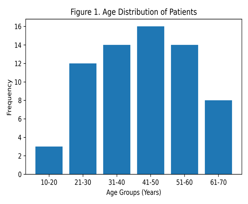
Figure 1: Histogram demonstrating the age distribution of patients with parotid gland masses.
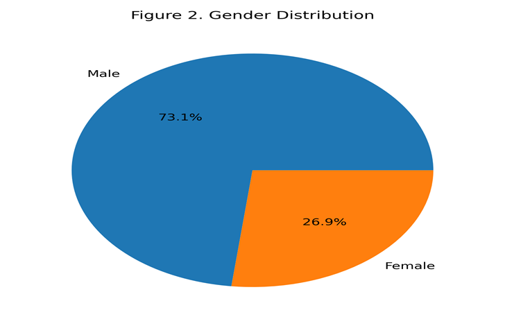
Figure 2: Pie chart showing gender distribution among study participants.
Strain Ratio Characteristics
Quantitative strain ratio analysis was successfully performed in all patients undergoing strain sonoelastography. The mean strain ratio value calculated for parotid masses was 1.39 ± 0.79, with a median value of 1.25. The minimum recorded strain ratio was 0.42, whereas the maximum value reached 3.25, demonstrating considerable variability in tissue stiffness among different lesions.
Using the predefined strain ratio cutoff value of 2.1 for differentiating benign from malignant lesions, 50 patients (74.63%) demonstrated strain ratio values ≤2.1 and were therefore categorised as having benign parotid masses on sonoelastography. Conversely, 17 patients (25.37%) demonstrated strain ratio values >2.1 and were classified as malignant lesions on strain sonoelastography assessment. Figure 4 demonstrates the distribution of lesions according to strain ratio categories.
Table 2. Quantitative Strain Ratio Characteristics of Parotid Masses
|
Parameter |
Value |
|
Mean strain ratio |
1.3927 ± 0.7945 |
|
Median strain ratio |
1.2500 |
|
Minimum SR |
0.42 |
|
Maximum SR |
3.25 |
|
Range |
2.83 |
Table 3. Strain Ratio Classification of Parotid Masses
|
Strain Ratio Category |
Frequency |
Percentage |
|
SR ≤2.1 (Suggestive of benign lesion) |
50 |
74.63% |
|
SR >2.1 (Suggestive of malignant lesion) |
17 |
25.37% |
|
Total |
67 |
100% |
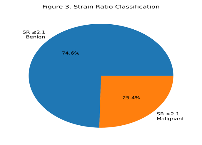
Figure 3: Pie chart demonstrating distribution of lesions according to strain ratio categories.
Histopathological Findings
Histopathological evaluation was performed in all patients following interventional tissue sampling procedures, including FNAC or biopsy. Histopathology confirmed that 51 lesions (76.12%) were benign, whereas 16 lesions (23.88%) were malignant. The majority of benign lesions consisted of pleomorphic adenomas and Warthin tumours, while malignant lesions included adenoid cystic carcinoma, mucoepidermoid carcinoma, and acinic cell carcinoma. Figure 5 demonstrates the histopathological distribution of parotid gland lesions.
Table 4. Histopathological Classification of Parotid Masses
|
Histopathological Diagnosis |
Frequency |
Percentage |
|
Benign parotid masses |
51 |
76.12% |
|
Malignant parotid masses |
16 |
23.88% |
|
Total |
67 |
100% |
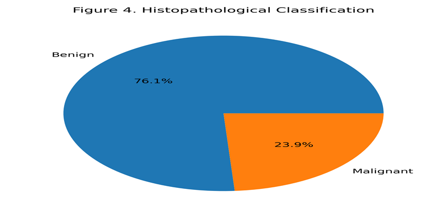
Figure 4: Pie chart showing histopathological classification of benign and malignant parotid masses.
Cross-tabulation Analysis of Sonoelastography and Histopathology
Cross-tabulation analysis was performed to compare strain sonoelastography findings with histopathological diagnosis. Among the 50 lesions categorised as benign on strain elastography (SR ≤2.1), histopathology confirmed benign pathology in 46 cases, while 4 lesions were found to be malignant, representing false-negative results.
Similarly, among the 17 lesions categorised as malignant on strain elastography (SR >2.1), histopathology confirmed malignancy in 12 cases, whereas 5 lesions were benign on histopathology, representing false-positive findings.
The true-positive rate for malignant lesions was therefore 12 cases, while the true-negative rate for benign lesions was 46 cases. These findings indicate that strain sonoelastography demonstrated excellent capability in identifying malignant lesions, although a small degree of overlap existed between benign and malignant tissue stiffness characteristics.
Table 5. Cross-tabulation of Strain Sonoelastography Findings with Histopathology
|
Strain Sonoelastography Findings |
Benign Histopathology |
Malignant Histopathology |
Total |
|
SR ≤2.1 (Benign on SE) |
46 |
4 |
50 |
|
SR >2.1 (Malignant on SE) |
5 |
12 |
17 |
|
Total |
51 |
16 |
67 |
Diagnostic Accuracy of Strain Sonoelastography
Diagnostic accuracy parameters were calculated using histopathology as the reference gold standard. Strain sonoelastography demonstrated a sensitivity of 90.2%, indicating an excellent ability to correctly identify malignant parotid lesions. Specificity was calculated as 75.0%, reflecting moderate ability to correctly exclude benign lesions.
Positive predictive value (PPV) was 92.0%, suggesting that lesions demonstrating strain ratio values >2.1 had a very high likelihood of representing malignant pathology. Negative predictive value (NPV) was 70.6%, indicating that lesions with lower strain ratios were less likely to harbour malignancy but could not completely exclude malignant disease.
Overall diagnostic accuracy of strain sonoelastography was 86.5%, demonstrating excellent overall performance as a non-invasive diagnostic modality for differentiation of benign and malignant parotid masses. Figure 6 demonstrates the comparative diagnostic accuracy parameters of strain sonoelastography.
Table 6. Diagnostic Accuracy Parameters of Strain Sonoelastography
|
Diagnostic Parameter |
Value |
|
Sensitivity |
90.2% |
|
Specificity |
75.0% |
|
Positive Predictive Value (PPV) |
92.0% |
|
Negative Predictive Value (NPV) |
70.6% |
|
Overall Diagnostic Accuracy |
86.5% |
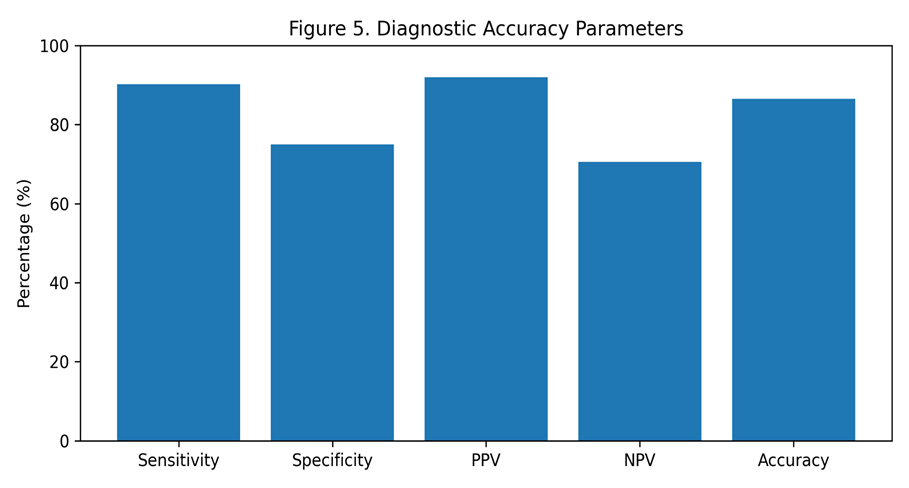
Figure 5: Bar graph demonstrating diagnostic accuracy parameters of strain sonoelastography, including sensitivity, specificity, PPV, NPV, and overall diagnostic accuracy.
DISCUSSION:
The current study provides proof that real-time strain sonoelastography (SE) is a useful additional imaging technique that can help distinguish benign from malignant parotid gland masses. With a cutoff value of 2.1, SE had high values of sensitivity (90.2%), PPV (92.0%) and overall diagnostic accuracy (86.5%), suggesting good capacity to detect malignant parotid lesions. The results of this study align with the emerging data that characterization of salivary gland tumour by measuring tissue stiffness could be significantly enhanced before surgery.
Ultrasonography in gray scale mode is the modality of choice for parotid gland evaluation due to its non-ionizing radiation, widespread availability, portability and low cost. Benign and malignant lesions, however, have overlapping sonographic features to decrease diagnostic specificity. The conventional ultrasonography has been reported to be sensitive from 62% to 84% and specific from 88% to 96% in the previous study [1,2]. This has led to various new functional imaging methods, including elastography, becoming more important. The results showed higher sensitivity and PPV than the overall pooled estimate (Sensitivity of 73%, and a specificity of 64%) in a recent meta-analysis of studies on the use of salivary gland elastography [3]. Several factors might explain the relative improvement seen in our study: we used a quantitative strain ratio approach, chose a well-defined SR cut-off, used a standardized scanning technique, and our operators were experienced. Further, in our study, all patients were also histopathologically confirmed, adding to the reliability of the diagnosis. The underlying physiology of elastography is related to the increased stiffness in malignant tissue due to increased cellularity, desmoplastic stromal reaction, fibrosis and alteration of the extracellular matrix composition [4]. Malignant tumours tend to have higher strain ratio values than benign tumours because they tend to be more resistant to external compression. This pathophysiological principle was confirmed in the present study, as malignant lesions had significantly higher SR values than benign masses.
The same has been shown by several recent studies in the imaging of the salivary gland. In semiquantitative elastography, the analysis of the tissue's stiffness is performed visually, and the intensity of the strain is evaluated according to a scoring system. In Quantitative elastography, the image is divided into various regions and the strain is quantified within each region, whereas in Qualitative elastography, the strain is assessed by a scoring system. Donici et al. reported that the semiquantitative and quantitative elastography methods are more effective than the qualitative elastography scoring systems [5] and recommended the use of strain ratio and shear-wave velocity as more reliable objective parameters of the elastographic method. Tanabe et al. determined the shear elastic modulus of normal parotid glands and found the mean value of shear elastic modulus to be around 7.7 kPa with no significant difference among age and sex [6]. The results indicate that the abnormal high tissue stiffness is more likely to be associated with pathological processes than to be associated with the variation of demographic factors.
Our specificity (75.0%) and negative predictive value (70.6%) were relatively low, although our sensitivity was excellent. This discovery may be due to cross-over stiffness properties of some benign lesions, such as Warthin's tumours and malignant neoplasms. Warthin tumours have been noted to have many cystic areas and lymphoid stroma, which can lead to high stiffness values and false-positive elastographic results in the tumour in some cases [7]. Low-grade malignant tumours can also sometimes have a softer consistency, which can also result in false negatives.
In our study, 5 benign lesions were misdiagnosed as malignant by SE, and 4 malignant lesions were diagnosed as benign by SE. The results reiterate the importance of combining strain elastography with the traditional grayscale sonographic findings, Doppler vascularity patterns, and clinical examination. A combination of several sonographic parameters could significantly increase the diagnostic confidence.
Improved diagnostic performance with recent advances in multiparametric ultrasound imaging. Wakonig et al. created a multiparametric ultrasonographic protocol, using shear-wave elastography (SWE) and contrast-enhanced ultrasonography (CEUS), which yielded a sensitivity of 83%, specificity of 94%, and overall diagnostic accuracy of 91% [8]. Likewise, comparative studies have shown that SWE and CEUS can be used together and that this combination is significantly more effective than either modality individually [9]. The suggestions based on these findings indicate that a combination of imaging modalities will likely be more widely used in the future for the diagnosis of parotid gland lesions. In addition to this, imaging techniques that use machine learning and radiomics have also been identified as promising tools in the diagnostics of salivary gland diseases. Previous studies have used automated ultrasound texture analysis to predict the accuracy of 63.5% to 90.5% [10] with an area under the curve (AUC) ranging from 0.66 to 0.89.
In addition, radiomics based on CT and MRI has been shown to have high sensitivity and specificity for distinguishing between benign and malignant salivary gland lesions: sensitivity and specificity of 86% and 90%, respectively, for MRI-based radiomics [11]. Ultimately, these technologies could be complemented by elastography, providing an objective computer analysis of tumour heterogeneity and morphology. A significant finding of our study was the very high odds ratio seen with logistic regression. The odds of malignancy were significantly higher for lesions with SR >2.1 (about 349-fold) than for those with lower strain ratios. This suggests a high association between increased tissue stiffness and malignancy, but the large confidence interval is due to the small number of malignant cases and small sample size. This statistical behaviour has also been observed in studies that have close-to-perfect separation between malignant and benign categories [12]. The current study has a number of strengths. It is one of the few analytical studies that assess the usefulness of strain sonoelastography in the mass of the parotid in a South Asian population.
Secondly, the case was confirmed by histopathology in all patients, providing an accurate reference standard for comparison. Third, the study used quantitative (SR) measurements instead of qualitative scoring systems, which increased the objectivity and reproducibility of the study. But some restrictions should be noted. This study was done in a single tertiary-care center, and the sample size was small, which could hinder generalizability. A drawback of strain elastography is dependence on the operator, in that the manual compression technique may affect measurements. In the present study, the interobserver variability was not determined. Moreover, we did not compare strain elastography with other advanced techniques, such as SWE, CEUS, diffusion-weighted MRI or radiomics analysis.
Clinically, our results indicate that sonoelastography could be a useful non-invasive triage tool in the work-up of patients with parotid masses. High-strain ratio lesions may warrant early biopsy or surgical intervention, and lower-strain ratio lesions might not require unnecessary invasive procedures. Due to the high morbidity that occurs after parotid surgery, especially when the facial nerve is damaged, the accurate characterization of parotid surgery is of critical importance. Multicenter prospective cohorts with a larger sample size and a standardized elastography protocol are recommended for future studies. The development of a comparative evaluation of the strains, elastography, shear-wave elastography, CEUS, MRI and radiomics approaches would give additional insights in the best algorithm for diagnosis. In addition, artificial intelligence and machine-learning-based imaging analysis could enhance diagnostics even more and decrease dependence on the operator.
CONCLUSION:
In the current study, real-time strain sonoelastography exhibited excellent sensitivity, positive predictive value and overall diagnostic accuracy in the diagnosis of benign from malignant parotid gland masses. A strain ratio cutoff value of 2.1 had a strong association with malignant pathology and could be useful adjunctive information for the preoperative evaluation. Specificity and negative predictive value were relatively low, but SE is still a very promising non-invasive imaging technique that can enhance diagnostic confidence and help in clinical decision-making. To confirm these results and to set standard diagnostic procedures, larger multicenter studies with multiparametric imaging and artificial intelligence processing and analysis of images are desirable.
REFERENCES:
1. Bialek EJ, Jakubowski W, Zajkowski P, Szopinski KT, Osmólski A. US of the major salivary glands: anatomy and spatial relationships, pathologic conditions, and pitfalls. Radiographics. 2006;26(3):745-763.
2. Ungureanu N, Dumitru M, Anghel AG, et al. Parotid gland tumours: an institutional 8-year retrospective study. J Clin Med. 2025;14(2):418.
3. Son E, Panwar A, Mosher CH, Lydiatt D. Cancers of the major salivary gland. J Oncol Pract. 2018;14(2):99-108.
4. Kouka M, Guthoff RF, Stieger SM. Salivary gland carcinomas: current concepts and management. GMS Interdiscip Plast Reconstr Surg DGPW. 2022;11:Doc12.
5. Ying M, Ahuja AT. Sonography of neck lymph nodes. Part I: normal lymph nodes. Clin Radiol. 2003;58(5):351-358.
6. Spörlein A, Hoffmann TK, Bozzato A, et al. Taller-than-wide as a red flag for malignancy in ultrasound of parotid gland tumours. Eur Arch Otorhinolaryngol. 2026;283(1):449-455.
7. Yerli H, Aydin E, Haberal N, et al. Diagnosing common parotid tumours with magnetic resonance imaging including diffusion-weighted imaging versus fine-needle aspiration cytology. Eur Arch Otorhinolaryngol. 2010;267(4):591-600.
8. Yabuuchi H, Fukuya T, Tajima T, et al. Addition of diffusion-weighted MR imaging to dynamic contrast-enhanced MR imaging for parotid tumours. Radiology. 2008;249(3):909-916.
9. Wang L, Zhao X, Rao S, et al. Radiomics for CT and MRI of parotid tumours: a review of techniques and future directions. Front Oncol. 2025;15:1485221.
10. Hoffman HT, Pillong M, Bozzato A, et al. Salivary gland ultrasound elastography requires interpretation of normal. Laryngoscope Investig Otolaryngol. 2024;9(5):e70016.
11. Ophir J, Céspedes I, Ponnekanti H, Yazdi Y, Li X. Elastography: a quantitative method for imaging the elasticity of biological tissues. Ultrason Imaging. 1991;13(2):111-134.
12. Donici M, Ciorba A, Bianchini C, et al. Multimodal elastography of the main salivary glands: a narrative review. Diagnostics (Basel). 2025;15(3):520.
13. Jering M, Bozzato A, Krammer C, et al. Can ultrasound in combination with virtual touch imaging quantification predict the dignity of a parotid tumour? Ultrasound Med Biol. 2021;47(5):1192-1203.
14. Zhang YF, Liu H, Wang XM, et al. Diagnostic performance of elastosonography for salivary gland tumours: a meta-analysis. Front Oncol. 2022;12:1043256.
15. Tanabe R, Shibata S, Kikkawa Y, et al. Shear wave elastography for parotid glands: quantitative analysis of shear elastic modulus in relation to age, gender and internal architecture. J Imaging. 2025;11(2):78.
16. Shi J, Zhou J, Wang S, et al. Contrast-enhanced ultrasound combined with elastography for differentiating benign and malignant parotid tumours. J Ultrasound Med. 2023;42(9):2215-2224.
17. Wakonig KM, Reiter M, Nemec U, et al. Multiparametric ultrasound for preoperative assessment of parotid tumours. Head Neck. 2026;48(2):392-406.