Study of Epidemiological & Clinical Profile of Chronic Obstructive Pulmonary Disease Patient at a Tertiary Care Hospital in Eastern Rajasthan.
- Manish Raj Pahadia , Professor, Department of General Medicine, Mahatma Gandhi Medical College & Hospital, Jaipur.
- Nirbhaydan Gadhavi , Junior Resident, Department of General Medicine, Mahatma Gandhi Medical College & Hospital, Jaipur.
- Mohit Agarwal , Associate Professor, Department of Pulmonary Medicine and Critical Care Medicine, NIMS University, Jaipur.
- Raman Sharma , Professor and Unit Head, Department of General Medicine, Mahatma Gandhi Medical College & Hospital, Jaipur.
- Mukesh K Sarna , Professor and Unit Head, Department of General Medicine, Mahatma Gandhi Medical College & Hospital, Jaipur.
- Akash Aggarwal , Junior Resident, Department of General Medicine, Mahatma Gandhi Medical College & Hospital, Jaipur.
- Saurabh Singh Soopa , Junior Resident, Department of General Medicine, Mahatma Gandhi Medical College & Hospital, Jaipur.
- Aman Bagariya , Junior Resident, Department of General Medicine, Mahatma Gandhi Medical College & Hospital, Jaipur.
- Robin Singh , Junior Resident, Department of General Medicine, Mahatma Gandhi Medical College & Hospital, Jaipur.
Article Information:
Abstract:
Background: Acute exacerbation of chronic obstructive pulmonary disease (AECOPD) is a major cause of morbidity, mortality, and healthcare utilisation. Comprehensive clinical profiling including inflammatory biomarkers, microbiological patterns, and cardiac complications in Indian AECOPD patients from Rajasthan remains limited. Aim: To study the clinical profile of patients with AECOPD at a tertiary care hospital in Rajasthan. Materials and Methods: This prospective observational study enrolled 105 patients with AECOPD admitted to the Department of General Medicine, MGMC&H, Jaipur, over 18 months. Demographic, clinical, laboratory (including NLR, PLR, CRP), spirometric, microbiological, radiological, and echocardiographic parameters were systematically assessed. Results: The mean age was 60.30 ± 10.20 years with male predominance (83.81%). Ever-smokers constituted 64.76% with mean 25.90 pack-years; biomass exposure in 27.62%. GOLD Group D predominated (77.14%). Mean FEV₁ was 43.10 ± 17.00% predicted; 62.86% had GOLD Stage III–IV. Infective causes precipitated 58.10% of exacerbations. Sputum culture was positive in 53.33%, with Klebsiella pneumoniae (30.36%) being the commonest isolate. Mean NLR was 6.40 ± 5.30, CRP 44.10 ± 25.20 mg/L. Type II respiratory failure was present in 38.10%. Pulmonary hypertension was detected in 59.05% and cor pulmonale in 39.05%. In-hospital mortality was 15.24%, all in patients with severe disease and respiratory failure. Conclusion: AECOPD patients at our centre present with advanced disease (77.14% GOLD Group D), high inflammatory burden, significant cardiac complications, and a microbiological profile dominated by Klebsiella pneumoniae—differing from Western literature where Haemophilus influenzae predominates. The 15.24% in-hospital mortality underscores the need for early recognition, aggressive management, and comprehensive post-discharge follow-up.
Keywords:
Article :
INTRODUCTION:
Chronic obstructive pulmonary disease (COPD) is a heterogeneous lung condition characterised by chronic respiratory symptoms due to abnormalities of the airways and/or alveoli, causing persistent, often progressive, airflow obstruction.1 It is the third leading cause of death globally, responsible for approximately 3.23 million deaths annually.2,3 India bears a disproportionate burden, with an estimated prevalence of 4.2–9.1% among adults above 30 years and the highest number of COPD deaths worldwide.4,5. Acute exacerbation of COPD (AECOPD) is defined as an event characterised by increased dyspnoea and/or cough and sputum that worsens in ≤14 days, often associated with increased local and systemic inflammation caused by infection, pollution, or other airway insults.1,6 AECOPD episodes accelerate lung function decline, impair quality of life, increase hospitalisation, and contribute significantly to mortality.7,8 Previous hospitalisation for AECOPD is an independent predictor of readmission (OR 3.1), with 6-month readmission rates exceeding 50%.9
The clinical profile of AECOPD is shaped by multiple factors including disease severity, comorbidities, precipitating cause, microbiological agents, inflammatory biomarkers, and the presence of cardiac complications such as pulmonary hypertension and cor pulmonale.10,11 Recent evidence has highlighted the prognostic value of readily available inflammatory biomarkers—particularly neutrophil-to-lymphocyte ratio (NLR), platelet-to-lymphocyte ratio (PLR), and C-reactive protein (CRP)—in risk stratification of AECOPD.12–14 The microbiological profile of AECOPD shows significant geographic variation, with Indian studies reporting a predominance of Gram-negative organisms, particularly Klebsiella pneumoniae, unlike Western literature where Haemophilus influenzae traditionally predominates.15,16 Despite the substantial COPD burden in Rajasthan—which has among the highest age-standardised COPD incidence rates in India4—comprehensive clinical profiling of AECOPD patients from this region remains limited. The present study was designed to systematically evaluate the clinical profile of AECOPD patients at a tertiary care hospital in Jaipur, encompassing demographic characteristics, risk factors, clinical presentation, inflammatory biomarkers, spirometric severity, microbiological profile, cardiac complications, and hospital outcomes.
MATERIALS AND METHODS:
Study Design: Prospective observational study.
Setting and Duration: Department of General Medicine, MGMC&H, Jaipur, Rajasthan, over 18 months. Approved by the IEC, MGMC&H, Jaipur. Conducted per Declaration of Helsinki and ICMR guidelines. Written informed consent obtained from all participants or legally authorised representatives.
Participants: 105 patients aged ≥40 years admitted with AECOPD (GOLD criteria). Exclusions: bronchial asthma, active pulmonary tuberculosis on treatment, bronchiectasis, interstitial lung disease, lung malignancy, and unstable cardiac conditions.
Data Collection: Demographics, smoking history (pack-years), biomass exposure, COPD duration, exacerbation history, comorbidities, clinical examination, laboratory investigations (CBC, DLC, NLR, PLR, ESR, CRP, ABG, RFT, LFT, RBS), spirometry (post-bronchodilator FEV₁, FEV₁/FVC), chest X-ray, ECG, 2D echocardiography (LVEF, PASP, cor pulmonale), sputum culture, and final outcome.
Statistical Analysis: SPSS; Mean ± SD for continuous, n (%) for categorical; Chi-square, Fisher’s exact, ANOVA; p<0.05 significant.
RESULTS:
Table 1: Demographic and Risk Factor Profile (N=105)
|
Characteristic |
Value |
|
Age Distribution |
|
|
Mean ± SD (Range) |
60.30 ± 10.20 (40–80) years |
|
40–49 years |
20 (19.05%) |
|
50–59 years |
26 (24.76%) |
|
60–69 years |
40 (38.10%) |
|
70–79 years |
17 (16.19%) |
|
≥80 years |
02 (01.90%) |
|
Gender (M:F = 5.2:1) |
|
|
Male |
88 (83.81%) |
|
Female |
17 (16.19%) |
|
Smoking History |
|
|
Ever-Smoker (Current + Ex) |
68 (64.76%) |
|
Mean Pack-Years |
25.90 ± 10.30 |
|
Cigarette / Bidi / Hookah |
33 (48.53%) / 30 (44.12%) / 05 (07.35%) |
|
Never-Smoker |
37 (35.24%) |
|
Biomass Fuel Exposure |
|
|
Present |
29 (27.62%) |
|
Mean Duration |
17.90 ± 8.50 years |
|
COPD Duration & Exacerbation History |
|
|
Mean COPD Duration |
7.70 ± 3.80 years |
|
Mean Exacerbations/Year |
2.40 ± 1.60 |
|
≥2 Exacerbations/Year |
77 (73.33%) |
|
≥2 Hospitalisations/Year |
44 (41.90%) |
The 60–69 years age group predominated (38.10%). The high proportion of ever-smokers (64.76%) with substantial pack-year burden, combined with 27.62% biomass exposure, reflects the dual risk factor profile characteristic of Rajasthan. The majority (73.33%) were frequent exacerbators.
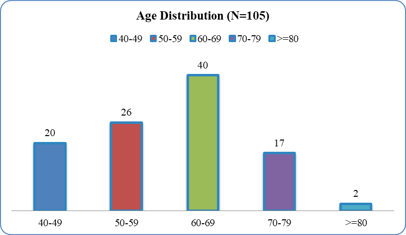
Figure 1: Age Distribution of Study Population (N=105)
Table 2: Clinical Presentation and Physical Examination (N=105)
|
Parameter |
n |
Percentage (%) |
|
Presenting Symptoms |
||
|
Dyspnoea |
104 |
99.05 |
|
Cough |
93 |
88.57 |
|
Increased Sputum |
87 |
82.86 |
|
Purulent Sputum |
66 |
62.86 |
|
Fever |
60 |
57.14 |
|
Pedal Oedema |
42 |
40.00 |
|
Physical Examination |
||
|
Cyanosis |
72 |
68.57 |
|
Accessory Muscle Use |
71 |
67.62 |
|
Pedal Oedema |
61 |
58.10 |
|
Raised JVP |
51 |
48.57 |
|
mMRC Dyspnoea Grade |
||
|
Grade 1 |
04 |
03.81 |
|
Grade 2 |
35 |
33.33 |
|
Grade 3 |
46 |
43.81 |
|
Grade 4 |
20 |
19.05 |
|
Comorbidities |
||
|
Hypertension |
49 |
46.67 |
|
Diabetes Mellitus |
37 |
35.24 |
|
Anaemia |
29 |
27.62 |
|
Coronary Artery Disease |
26 |
24.76 |
Dyspnoea was near-universal (99.05%). mMRC Grade 3–4 in 62.86% indicates severe functional limitation. The high prevalence of cyanosis (68.57%) and accessory muscle use (67.62%) reflects significant respiratory distress. Hypertension (46.67%) and diabetes (35.24%) were the commonest comorbidities.
Table 3: Laboratory and Inflammatory Parameters (N=105)
|
Parameter |
Mean ± SD |
Range |
|
Haematological |
||
|
Haemoglobin (g/dL) |
11.90 ± 2.50 |
6.50–17.00 |
|
TLC (/mm³) |
13847 ± 4094 |
6753–25796 |
|
Neutrophils (%) |
75.60 ± 9.60 |
55–95 |
|
Inflammatory Biomarkers |
||
|
NLR |
6.40 ± 5.30 |
1.57–19.00 |
|
PLR |
137.00 ± 130.10 |
21.20–565.70 |
|
ESR (mm/hr) |
33.60 ± 15.90 |
5–71 |
|
CRP (mg/L) |
44.10 ± 25.20 |
3.20–123.00 |
|
Arterial Blood Gas |
||
|
pH |
7.37 ± 0.06 |
7.25–7.48 |
|
PaO₂ (mmHg) |
57.70 ± 11.80 |
35–85 |
|
PaCO₂ (mmHg) |
52.90 ± 12.90 |
32–88 |
|
HCO₃ (mEq/L) |
25.10 ± 4.80 |
16–35 |
Neutrophilic leucocytosis was characteristic. Inflammatory biomarkers were uniformly elevated: mean NLR 6.40 (above the prognostic threshold of 5.67 reported by Chen et al.), CRP 44.10 mg/L (above the 30 mg/L threshold by Patil et al.). Type II respiratory failure was present in 40 (38.10%); overall respiratory failure in 58 (55.24%).
Table 4: Spirometric Severity, GOLD Classification, and Precipitating Factors (N=105)
|
Parameter |
n (%) / Value |
|
Spirometric Values |
|
|
FEV₁ (% predicted) — Mean ± SD |
43.10 ± 17.00 (Range: 13–86) |
|
FEV₁/FVC Ratio |
0.55 ± 0.08 |
|
GOLD Spirometric Stage |
|
|
Stage I (Mild, ≥80%) |
03 (02.86%) |
|
Stage II (Moderate, 50–79%) |
36 (34.29%) |
|
Stage III (Severe, 30–49%) |
45 (42.86%) |
|
Stage IV (Very Severe, <30%) |
21 (20.00%) |
|
GOLD ABCD Group |
|
|
Group A |
02 (01.90%) |
|
Group B |
20 (19.05%) |
|
Group C |
02 (01.90%) |
|
Group D |
81 (77.14%) |
|
Cause of Exacerbation |
|
|
Acute Bronchitis |
23 (21.90%) |
|
Pneumonia |
21 (20.00%) |
|
Pulmonary Tuberculosis |
11 (10.48%) |
|
Non-Compliance |
08 (07.62%) |
|
Heart Failure |
07 (06.67%) |
|
URTI |
06 (05.71%) |
|
CLD with Pleural Effusion |
06 (05.71%) |
|
CKD |
05 (04.76%) |
|
GI Infection |
02 (01.90%) |
|
Miscellaneous |
16 (15.24%) |
GOLD Stage III–IV accounted for 62.86%, and 77.14% were Group D. Infective causes collectively precipitated 58.10% of exacerbations. Pulmonary TB (10.48%) as a precipitant reflects the high TB burden in India.
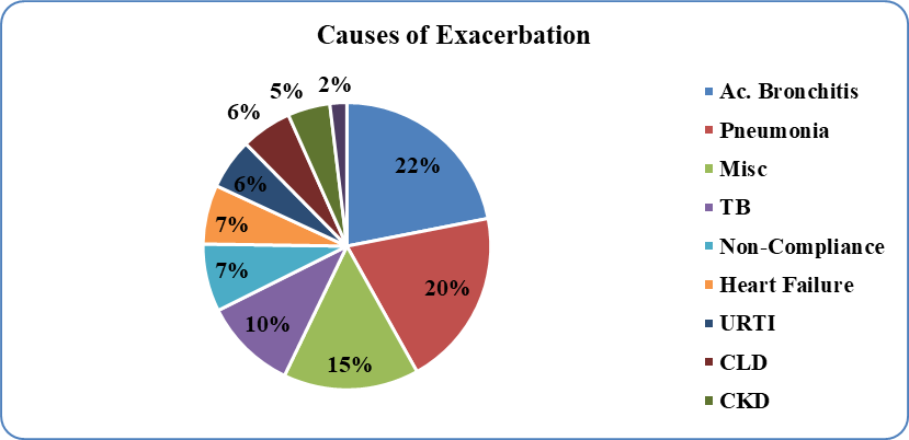
Figure 2: Causes of Acute Exacerbation (N=105)
Table 5: Cardiac Evaluation and Pulmonary Hypertension (N=105)
|
Parameter |
n (%) / Value |
|
Chest X-Ray Findings |
|
|
Hyperinflation |
69 (65.71%) |
|
Prominent Pulmonary Artery |
57 (54.29%) |
|
Cardiomegaly |
54 (51.43%) |
|
Consolidation |
50 (47.62%) |
|
Bullae |
26 (24.76%) |
|
ECG Findings |
|
|
P-Pulmonale |
54 (51.43%) |
|
Right Ventricular Hypertrophy |
52 (49.52%) |
|
Atrial Fibrillation |
19 (18.10%) |
|
Echocardiography |
|
|
Mean LVEF (%) |
53.20 ± 8.40 (Range: 25–70) |
|
Mean PASP (mmHg) |
39.90 ± 16.50 (Range: 16–77) |
|
Pulmonary Hypertension (Any) |
62 (59.05%) |
|
Mild / Moderate / Severe |
20 (19.05%) / 27 (25.71%) / 15 (14.29%) |
|
Cor Pulmonale |
41 (39.05%) |
Pulmonary hypertension was present in 59.05% and cor pulmonale in 39.05%, reflecting advanced cardiopulmonary disease. P-pulmonale (51.43%) and RVH (49.52%) on ECG corroborated the echocardiographic findings.
D
Table 6: Sputum Culture and Microbiological Profile (N=105)
|
Parameter |
n |
Percentage (%) |
|
Culture Result |
||
|
Culture Positive |
56 |
53.33 |
|
No Growth |
49 |
46.67 |
|
Organisms Isolated (n=56) |
||
|
Klebsiella pneumoniae |
17 |
30.36 |
|
Streptococcus pneumoniae |
10 |
17.86 |
|
Moraxella catarrhalis |
09 |
16.07 |
|
Haemophilus influenzae |
06 |
10.71 |
|
Pseudomonas aeruginosa |
05 |
08.93 |
|
Acinetobacter species |
05 |
08.93 |
|
Escherichia coli |
02 |
03.57 |
|
Mycobacterium tuberculosis |
02 |
03.57 |
53.33% culture positivity rate is consistent with Groenewegen and Wouters (50%). Klebsiella pneumoniae predominance (30.36%) differs from Western literature (H. influenzae) and is consistent with Indian studies by Sharma et al. Gram-negative organisms predominated overall.
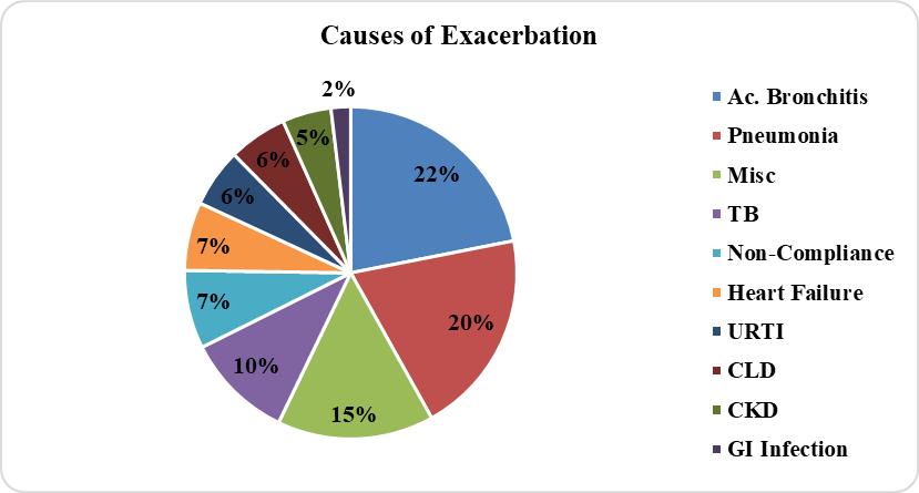
Figure 3: Sputum Culture Isolates (n=56)
Table 7: Hospital Course and Outcome (N=105)
|
Parameter |
n (%) / Value |
|
Ventilatory Support |
|
|
Non-Invasive Ventilation |
46 (43.81%) — Mean 2.60 ± 1.50 days |
|
Invasive Mechanical Ventilation |
31 (29.52%) — Mean 3.30 ± 1.70 days |
|
Hospital Stay |
|
|
Mean Hospital Stay (days) |
10.20 ± 3.90 (Range: 3–20) |
|
ICU Admission |
36 (34.29%) — Mean 4.00 ± 1.80 days |
|
Final Outcome |
|
|
Improved and Discharged |
81 (77.14%) |
|
Death |
16 (15.24%) |
|
DAMA |
06 (05.71%) |
|
Referred to Higher Centre |
02 (01.90%) |
In-hospital mortality was 15.24%, comparable to Kumar and Choubey (17%) and Suresh et al. (18.3%). All fatalities occurred in patients with respiratory failure requiring ventilatory support. The 29.52% invasive ventilation rate and 34.29% ICU admission rate reflect the severity of the admitted cohort.
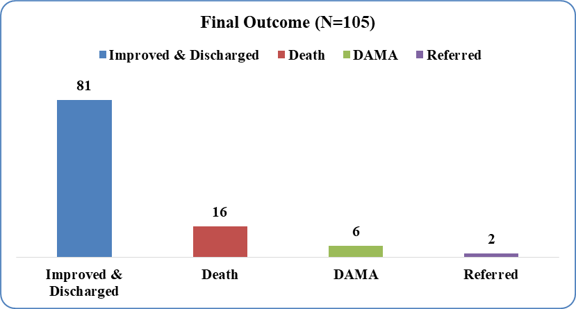
Figure 4: Final Outcome (N=105)
DISCUSSION:
Demographic Profile
The mean age of 60.30 ± 10.20 years is consistent with Patil et al.17 (62.1 years), Kumar and Choubey18 (62.4 years), and Ramakrishna et al.19 (64.2 years). The marginally lower mean age may reflect earlier disease onset in Rajasthan due to biomass exposure, bidi smoking, and occupational dust. Male predominance (83.81%) aligns with Patil et al.17 (88%), Kumar and Choubey18 (78.6%), and Suresh et al.20 (82.5%). The 27.62% biomass exposure rate, predominantly in females, is consistent with Sharma et al.21 (34.6% in 2,194 Indian COPD patients), who identified a distinct biomass-COPD phenotype with more small airway disease and earlier pulmonary hypertension.
Inflammatory Biomarkers
The elevated NLR (6.40 ± 5.30) exceeds the prognostic threshold of 5.67 reported by Chen et al.12 (AUC 0.801 for 28-day mortality). Jain et al.13 from Bikaner, Rajasthan, reported NLR 5.72 vs 3.77 in AECOPD vs stable COPD (AUC 0.782). Al-Lawati et al.14 documented NLR 6.84 and CRP 48.6 mg/L, with combined AUC 0.912. Wang et al.22 demonstrated NLR ≥4.43 independently predicted readmission (HR 1.78). Srinivas et al.23 showed progressive NLR increase across mild (4.12), moderate (6.78), and severe (9.56) AECOPD. The mean CRP of 44.10 mg/L exceeds the 30 mg/L threshold identified by Patil et al.24 as predicting adverse outcomes (OR 2.89). These findings support NLR and CRP as readily available prognostic tools in AECOPD.
Respiratory Failure and Pulmonary Hypertension
Respiratory failure in 55.24% with Type II predominance (38.10%) is consistent with the pathophysiology of severe COPD. Patil et al.17 documented respiratory failure in 33.8% with hypercapnia as an independent mortality predictor. Pulmonary hypertension in 59.05% and cor pulmonale in 39.05% exceed the ACURE registry10 (25.28% PHD), likely reflecting the advanced disease profile (62.86% GOLD III–IV) and biomass exposure. Kumar and Choubey18 reported cor pulmonale in 42.1%, comparable to our findings.
Microbiological Profile
The 53.33% culture positivity with Klebsiella pneumoniae predominance (30.36%) contrasts with Western literature where H. influenzae predominates. This mirrors Sharma et al.15 (K. pneumoniae 38.6%) and reflects the geographic variation in AECOPD bacteriology. Groenewegen and Wouters16 reported 50% positivity with H. influenzae (45%). This has direct implications for empirical antibiotic selection in Indian AECOPD patients—favouring agents with Gram-negative coverage.
Outcomes and Mortality
The 15.24% in-hospital mortality is concordant with Kumar and Choubey18 (17%) and Suresh et al.20 (18.3%). Liu et al.25 in their meta-analysis of 37 studies identified age (OR 1.45), pneumonia (OR 2.12), acidosis (OR 2.78), and mechanical ventilation (OR 4.23) as mortality predictors—all highly prevalent in our cohort. The mean hospital stay (10.20 days) is comparable to Sharma et al.26 (10.9 days). The 41.90% with ≥2 prior hospitalisations and 39.05% with cor pulmonale predict high readmission risk.
CONCLUSION:
The clinical profile of AECOPD patients at our tertiary care centre in Rajasthan is characterised by advanced disease severity (77.14% GOLD Group D, 62.86% GOLD Stage III–IV), high inflammatory burden (mean NLR 6.40, CRP 44.10 mg/L), significant cardiac complications (59.05% pulmonary hypertension, 39.05% cor pulmonale), and a distinctive microbiological profile dominated by Klebsiella pneumoniae (30.36%). The in-hospital mortality of 15.24% underscores the need for early recognition, aggressive management including appropriate antibiotic selection guided by local microbiological patterns, biomarker-based risk stratification using NLR and CRP, echocardiographic assessment for pulmonary hypertension and cor pulmonale, and comprehensive post-discharge follow-up to reduce readmissions.
LIMITATIONS
1. Single-centre design.
2. Viral diagnostics not available; viral triggers may be underestimated.
3. GOLD ABCD classification based on recall of pre-exacerbation symptoms.
REFERENCES:
1. Agustí A, Celli BR, Criner GJ, et al. Global Initiative for Chronic Obstructive Lung Disease 2023 Report: GOLD Executive Summary. Eur Respir J. 2023;61(4):2300239.
2. Adeloye D, Song P, Zhu Y, et al. Global prevalence of and risk factors for COPD in 2019: a systematic review. Lancet Respir Med. 2022;10(5):447-58.
3. Al Wachami N, Guennouni M, Iderdar Y, et al. Estimating global prevalence of COPD: a systematic review. BMC Public Health. 2024;24(1):297.
4. Salvi S, Kumar GA, Dhaliwal RS, et al. Burden of chronic respiratory diseases in India: GBD 1990-2016. Lancet Glob Health. 2018;6(12):e1363-74.
5. Verma A, Gudi N, Yadav UN, et al. Prevalence of COPD among population above 30 years in India: systematic review. J Glob Health. 2021;11:04038.
6. Ko FWS, Chan KP, Hui DSC. Acute exacerbation of COPD. Respirology. 2020;25(7):719-28.
7. Hurst JR, Vestbo J, Anzueto A, et al. Susceptibility to exacerbation in COPD. N Engl J Med. 2010;363(12):1128-38.
8. Rothnie KJ, Müllerová H, Smeeth L, Quint JK. Natural history of COPD exacerbations in general practice. Am J Respir Crit Care Med. 2018;198(4):464-71.
9. Dewan NA, Rafique S, Kanwar B, et al. Acute exacerbation of COPD: factors associated with poor outcomes. Chest. 2000;117(3):662-71.
10. Sun Y, Cai BQ, Wang XJ, et al. Clinical features and outcomes of AECOPD with PHD: ACURE registry. Int J Chron Obstruct Pulmon Dis. 2021;16:2605-16.
11. Qian Y, Cai C, Sun M, et al. Factors associated with AECOPD: a review. Int J Chron Obstruct Pulmon Dis. 2023;18:2707-23.
12. Chen Y, Luo D, Zhang K, et al. NLR and PLR as predictors of 28-day mortality in AECOPD. Int Immunopharmacol. 2021;94:107476.
13. Jain A, Sharma S, Gupta R. NLR in AECOPD: a hospital-based study from Rajasthan. Indian J Allergy Asthma Immunol. 2022;36(1):23-8.
14. Al-Lawati A, Al-Rawas O, Al-Riyami B. NLR and CRP in AECOPD: diagnostic accuracy study. Lung. 2023;201(3):287-95.
15. Sharma P, Agarwal SK, Sharma N. Bacteriological profile of AECOPD in India. Indian J Chest Dis Allied Sci. 2015;57(2):93-6.
16. Groenewegen KH, Wouters EF. Bacterial infections in patients requiring admission for AECOPD. Respir Med. 2003;97(7):770-7.
17. Patil SP, Krishnan JA, Lechtzin N, Diette GB. In-hospital mortality following AECOPD. Arch Intern Med. 2003;163(10):1180-6.
18. Kumar H, Choubey S. Clinical profile of AECOPD at a tertiary care hospital in India. J Med Sci Clin Res. 2018;6(7):467-74.
19. Ramakrishna R, Kumar S, Patil M. Predictors of outcome in hospitalised AECOPD. Indian J Respir Care. 2023;12(1):42-8.
20. Suresh V, Mohan S, Raghavan R. Clinical profile and outcomes of AECOPD: a tertiary care study. Lung India. 2024;41(3):178-85.
21. Sharma BB, Singh V, Sharma S, et al. Multicenter phenotyping of COPD patients in India. Lung India. 2022;39(6):518-27.
22. Wang H, Zhang L, Chen Y. NLR as predictor of readmission in AECOPD: 8-year longitudinal study. BMC Pulm Med. 2023;23:156.
23. Srinivas K, Reddy P, Mohan A. Inflammatory biomarkers and AECOPD severity: a cross-sectional study. J Clin Diagn Res. 2025;19(1):OC01-5.
24. Patil R, Desai A, Shah P. CRP and NLR as predictors of adverse outcomes in AECOPD. Indian J Chest Dis Allied Sci. 2025;67(1):12-8.
25. Liu J, Zhang W, Chen Y. Predictors of mortality in hospitalised AECOPD: systematic review and meta-analysis. Respir Res. 2023;24(1):165.
26. Sharma S, Gupta A, Bansal P. Predictors of prolonged hospitalisation in AECOPD. Lung India. 2024;41(5):386-93.