Spectrum of Cardiac Dysfunction in Chronic Liver Disease and Its Association with Child–Pugh Classification.
- Omkaram Siddhardha Varma , Senior Resident, Department of General Medicine, ESIC Medical College & Hospital, Hyderabad, Telangana, India.
- Dinesh Poonia , Classified Specialist, Department of General Medicine, 167 Military Hospital, Pathankot, Punjab, India.
- Chennakesavulu Dara , Associate Professor, Department of General Medicine, ESIC Medical College & Hospital, Hyderabad, Telangana, India.
- Veershetty Pasari , Senior Resident, Department of General Medicine, ESIC Medical College & Hospital, Hyderabad, Telangana, India.
- Arati Ambrish Shahade , Consultant Physician & Diabetologist, Department of General Medicine, Shahade Hospital, Pune, Maharashtra, India.
Article Information:
Abstract:
Background: Chronic liver disease (CLD) exerts pathological effects on multiple organ systems, including the cardiovascular system, which often remain clinically unnoticed. In many patients with CLD, cardiovascular complications develop in a subclinical manner and become evident only under stressful conditions. Therefore, early detection of cirrhotic cardiomyopathy using electrocardiography (ECG) and two-dimensional echocardiography (2D-ECHO) may help reduce morbidity and mortality in these patients. Objectives: To identify cardiac dysfunction in patients with chronic liver disease using electrocardiography and echocardiography. To correlate cardiac abnormalities with the severity of chronic liver disease based on the Child–Pugh scoring system. Methodology: This multicenter study enrolled 100 patients with chronic liver disease. Clinical data were recorded using a standardized proforma. All participants underwent ECG and echocardiographic assessment, and liver disease severity was graded using the Child–Pugh classification. Cardiac abnormalities were evaluated and correlated with the severity of underlying liver disease.. Results: Among the 100 patients studied, 81% were males and 78% had a history of alcoholism. Abnormal ECG findings were observed in 57% of patients, with QT prolongation being the most common abnormality, present in 29% of cases, and showing correlation with the severity of liver disease. Elevated pro-BNP levels were observed in 77% of patients with significant cardiac dysfunction. Most patients belonged to Child–Pugh class C. Echocardiographic evaluation was normal in 48% of patients, while diastolic dysfunction was the most common abnormal finding, seen in 44% of cases, and was positively correlated with the severity of liver disease. Conclusion: QT prolongation was the most common ECG abnormality observed in patients with chronic liver disease. Diastolic dysfunction was the most frequent echocardiographic abnormality and showed a strong correlation with the severity of liver disease.
Keywords:
Article :
INTRODUCTION:
Chronic liver disease (CLD) is a major global health problem associated with significant morbidity and mortality. In addition to hepatic dysfunction, CLD affects multiple organ systems, including the cardiovascular system. Cirrhotic cardiomyopathy is characterized by impaired cardiac contractility, diastolic dysfunction, and electrophysiological abnormalities in the absence of pre-existing heart disease. These cardiac changes often remain asymptomatic at rest and become clinically evident only during physiological stress, infections, or invasive procedures such as liver transplantation(1).
Electrocardiographic abnormalities, particularly prolonged QT interval, and echocardiographic evidence of diastolic dysfunction are commonly reported in patients with advanced liver disease(2)Early recognition of these abnormalities is important because they contribute to increased perioperative risk, reduced exercise tolerance, and poor clinical outcomes. The severity of liver dysfunction assessed by the Child–Pugh scoring system has been shown to correlate with the degree of cardiovascular impairment (3). Therefore, evaluation of cardiac abnormalities using electrocardiography and echocardiography may facilitate timely diagnosis and improve management strategies in patients with chronic liver disease.
MATERIALS AND METHODS:
A multicenter cross-sectional observational study was conducted over 18 months, from December 2020 to June 2022, at ESIC Medical College and Hospital, Army Hospital Pathankot, and Shahade Hospial, Pune. A total of 100 patients with chronic liver disease were enrolled in the study. Patients aged above 18 years with clinical and/or radiological evidence of chronic liver disease or cirrhosis of any etiology and disease duration greater than 26 weeks were included. Patients with pre-existing cardiac disorders such as congenital heart disease, ischemic heart disease, congestive cardiac failure, or comorbid conditions including diabetes mellitus and hypertension were excluded from the study. Written informed consent was obtained from all participants prior to enrolment. Detailed clinical evaluation was performed in each patient, including assessment for signs of chronic liver disease, hepatic encephalopathy, blood pressure measurement, and comprehensive cardiovascular examination.
All participants underwent relevant laboratory investigations, including complete blood count, liver function tests, renal function tests, PT/INR, aPTT, viral markers (HBsAg and anti-HCV), and serum pro-BNP estimation in selected cases. Ultrasonography of the abdomen, electrocardiography (ECG), and two-dimensional echocardiography (2D-ECHO) were performed to evaluate cardiovascular abnormalities with cardiology consultation. The severity of liver disease was graded using the Child–Pugh scoring system. Statistical analysis was carried out using SPSS version 18.0 and R software version 3.2.2. Continuous variables were expressed as mean ± standard deviation, while categorical variables were presented as frequencies and percentages. ANOVA, Chi-square test, and Fisher’s exact test were applied wherever appropriate, and a p-value of ≤0.05 was considered statistically significant.
RESULTS:
A total of 100 patients with chronic liver disease (CLD) were included in the study and underwent detailed cardiac evaluation to assess cardiovascular abnormalities and their correlation with the severity of liver disease. The majority of patients belonged to the age group of 41–50 years, followed by 30–40 years and 51–60 years, together accounting for 82% of the study population. The mean age of the participants was 43.22 ± 10.43 years.
Male predominance was observed, with males constituting 81% of the study population, while females accounted for 19%. The most common clinical presentations were abdominal distension (98%) and lower limb edema (97%). Jaundice was present in 64% of patients, gastrointestinal bleeding in 42%, altered sensorium in 24%, and breathlessness in 13% of cases. A history of alcoholism was observed in 78% of patients, and more than half of them (54%) reported alcohol consumption for over 10 years. Laboratory evaluation revealed that 44% of patients had serum bilirubin levels below 2.5 mg/dL, whereas 42% had bilirubin levels greater than 5 mg/dL. Hypoalbuminemia was common, with 76% of patients having serum albumin levels between 1–2.5 g/dL. Coagulopathy with INR >1.5 was noted in 66% of patients.
Serum pro-BNP levels were assessed in 90 patients, among whom 77.8% showed elevated values. Most patients with raised pro-BNP levels belonged to Child–Pugh class C and demonstrated associated echocardiographic evidence of diastolic dysfunction. Electrocardiographic abnormalities (Table.1) were identified in 57% of patients. QT interval prolongation was the most frequent ECG abnormality, observed in 29% of patients, followed by right bundle branch block (RBBB) in 6% of cases. The majority of study participants (74%) were categorized under Child–Pugh class C, whereas only 4% belonged to Child–Pugh class A. Severe liver disease (Child–Pugh class C) was particularly common among patients aged 41–50 years.
Table 1: Association between Severity of CLD and ECG changes
|
|
Child Pugh class |
Total |
||
|
A |
B |
C |
||
|
Normal |
4 |
18 |
21 |
43 |
|
Sinus Tachycardia |
0 |
2 |
20 |
22 |
|
RBBB |
0 |
0 |
6 |
6 |
|
QT Prolongation |
0 |
2 |
27 |
29 |
|
Total |
4 |
22 |
74 |
100 |
|
Chi square test= 25.51, p=0.0001* Statistically significant |
||||
Echocardiographic evaluation revealed normal findings (Table.2) in 48% of patients, while diastolic dysfunction was the most common abnormality, detected in 44% of cases. Among patients with QT prolongation, 93.1% belonged to Child–Pugh class C. Similarly, a significant proportion of patients with diastolic dysfunction were also categorized under Child–Pugh class C, indicating a strong association between cardiac dysfunction and the severity of liver disease.
Table 2: Echocardiographic manifestations in CLD
|
|
Frequency |
Percentage |
|
Normal |
48 |
48% |
|
Diastolic dysfunction |
44 |
44% |
|
Pulmonary HTN |
3 |
3% |
|
Dilated Cardiomyopathy |
1 |
1% |
|
Systolic and Diastolic dysfunction |
4 |
4% |
Echocardiographic abnormalities increased significantly with advancing liver disease severity. Most patients with Child–Pugh Class C had cardiac dysfunction, predominantly diastolic dysfunction (41%), while normal echocardiographic findings were more common in Classes A and B. This association was statistically significant (χ² = 23.48, p = 0.001), indicating worsening cardiac involvement with progressive cirrhosis (Graph.1).
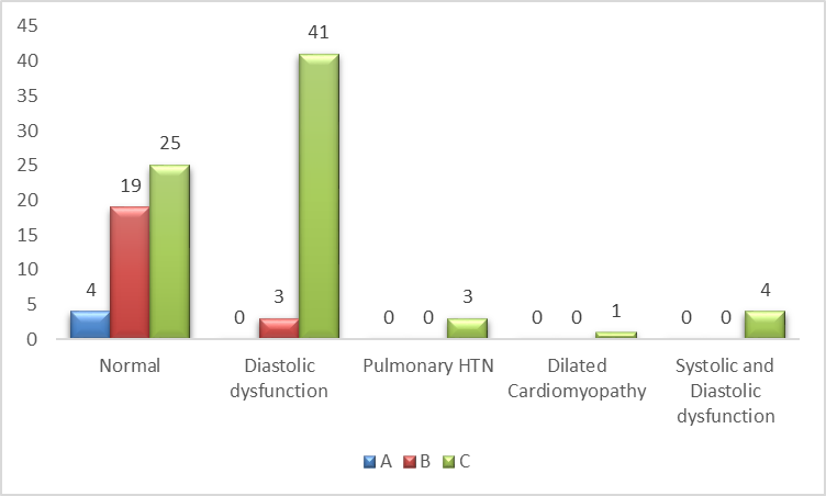
Graph 1: Correlation of severity of liver disease and Echocardiographic manifestations
These findings suggest that both electrocardiographic and echocardiographic abnormalities become increasingly prevalent with advancing hepatic dysfunction.
DISCUSSION:
Cirrhotic cardiomyopathy (CCM) is an increasingly recognized cardiovascular complication of chronic liver disease that often remains underdiagnosed because patients are usually asymptomatic at rest. It is characterized by impaired cardiac contractility during stress, diastolic dysfunction, and electrophysiological abnormalities in the absence of primary heart disease. Early diagnosis is important because undetected CCM can increase perioperative and post-transplant morbidity and mortality in patients undergoing liver transplantation.
In the present study, most patients belonged to the 41–50 years age group, which is comparable to findings reported by Kazankov et al., Kim et al., and Fischer et al., where the mean age ranged from 50 to 53 years (4). Increasing age was associated with higher Child-Pugh scores, indicating greater severity of liver disease and increased risk of cardiac dysfunction.
A marked male predominance (81%) was observed, likely due to the high prevalence of alcohol-related liver disease. Similar observations were reported in studies by Rabie et al. and other investigators (5,6). Alcoholism was the most common etiology of cirrhosis in the present study, accounting for 78% of cases, while viral hepatitis was the second most common cause. Chronic alcohol exposure may contribute to both hepatic and myocardial injury, thereby increasing the risk of CCM (7).
Most patients in this study belonged to Child-Pugh class C, suggesting advanced liver dysfunction. Similar studies have demonstrated a strong association between worsening liver disease and cardiac abnormalities, particularly QTc prolongation. Elevated pro-BNP levels were observed in the majority of patients, consistent with findings by Henriksen et al (8). Increased BNP secretion in cirrhosis is related to hyperdynamic circulation, ventricular stress, and neurohormonal activation.
Electrocardiographic abnormalities were present in more than half of the patients, with QT prolongation being the most common finding. Most patients with prolonged QT interval belonged to Child-Pugh class C, indicating a close relationship between electrophysiological abnormalities and severity of cirrhosis (9). Sinus tachycardia and right bundle branch block were also observed. Previous studies have suggested that alcohol-related cirrhosis is more frequently associated with QT prolongation compared to viral etiologies.
Echocardiographic evaluation showed diastolic dysfunction as the most common manifestation of CCM, affecting 44% of patients. Similar findings were reported by Mihalovici et al. and Puncho et al (10). Diastolic dysfunction was predominantly seen in Child-Pugh class C patients, suggesting that worsening hepatic dysfunction parallels deterioration in cardiac performance. Systolic dysfunction was less common and usually becomes evident only during stress conditions (11).
Overall, the present study highlights the high prevalence of cardiac abnormalities among patients with advanced liver cirrhosis, particularly in alcohol-related disease and higher Child-Pugh classes. Routine cardiac evaluation using ECG, echocardiography, and biomarkers such as BNP and NT-proBNP may facilitate early diagnosis and improve clinical outcomes, especially in patients being considered for liver transplantation (12, 13).
CONCLUSION:
Cardiac abnormalities are common in chronic liver disease and increase with the severity of cirrhosis. Diastolic dysfunction was the predominant echocardiographic finding, while QT interval prolongation was the most frequent ECG abnormality. Elevated serum pro-BNP levels correlated with disease progression, supporting its value as a marker of cardiac involvement. Routine cardiovascular screening with ECG, echocardiography, and biomarkers may facilitate early detection of cirrhotic cardiomyopathy, improve risk assessment, and optimize patient outcomes, particularly in advanced liver disease and liver transplantation candidates.
REFERENCES:
1. Møller S, Henriksen JH. Cirrhotic cardiomyopathy: a pathophysiological review of circulatory dysfunction in liver disease. Heart. 2002;87(1):9–15.
2. Zambruni A, Trevisani F, Caraceni P, Bernardi M. Cardiac electrophysiological abnormalities in patients with cirrhosis. J Hepatol. 2006;44(5):994–1002.
3. Kazankov K, Holland-Fischer P, Andersen NH, et al. Resting myocardial dysfunction in cirrhosis quantified by tissue Doppler imaging. Liver Int. 2011;31(4):534–540.
4. Kim MY, Baik SK, Won CS, Park HJ, Jeon HK, Hong HI, et al. Dobutamine stress echocardiography for evaluating cirrhotic cardiomyopathy in liver cirrhosis. Korean J Hepatol. 2010;16(4):376-82.
5. El-Shabrawi MH, Omran S, Wageeh S, Isa M, Okasha S, Mohsen NA, et al. (99m)Technetium-macroaggregated albumin perfusion lung scan versus contrast enhanced echocardiography in the diagnosis of the hepatopulmonary syndrome in children with chronic liver disease. Eur J Gastroenterol Hepatol. 2010;22(8):1006-12.
6. Rabie RN, Cazzaniga M, Salerno F, Wong F. The use of E/A ratio as a predictor of outcome in cirrhotic patients treated with transjugular intrahepatic portosystemic shunt. Am J Gastroenterol. 2009;104(10):2458-66.
7. Hammami R, Boudabbous M, Jdidi J, Trabelsi F, Mroua F, Kallel R, Amouri A, AbidD,Tahri N, Abid L, Kammoun S. Cirrhotic cardiomyopathy: is there any correlation between the stage of cardiac impairment and the severity of liver disease? Libyan J Med. 2017 Dec;12(1):1283162
8. Henriksen JH, Gøtze JP, Fuglsang S, Christensen E, Bendtsen F, Møller S. Increased circulating pro-brain natriuretic peptide (proBNP) and brain natriuretic peptide (BNP) in patients with cirrhosis: relation to cardiovascular dysfunction and severity of disease. Gut. 2003 Oct;52((10)):1511–7.
9. L. Pourafkari, et al., Electrocardiographic findings in hepatic cirrhosis and their association with the severity of disease, Cor et Vasa 59 (2017) e105–e113.
10. Mihailovici AR, Donoiu I, Gheonea DI, Mirea O, Târtea GC, Buşe M, Calborean V, Obleagă C, Pădureanu V, Istrătoaie O. NT-proBNP and Echocardiographic Parameters in Liver Cirrhosis - Correlations with Disease Severity. Med Princ Pract. 2019 Apr 16;28(5):432–41. doi: 10.1159/000499930. Epub ahead of print. PMID: 30995644; PMCID: PMC6771067.
11. Punekar P, Thakur DK. Echocardiographic changes in chronic liver disease. International Journal of Contemporary Medical Research. 2018;5(3):C01-04.
12. Ripoll C, Catalina MV, Yotti R, Olmedilla L, Pérez-Peña J, Lo Iacono O, et al. Cardiacdysfunction during liver transplanta- tion: incidence and preoperative predictors. Transplantation 2008; 85: 1766–1772, doi: 10.1097/TP.0b013e318172c936.
13. Ruiz-del-Arbol, Achecar L, Serradilla R, Rodriguez Gandia MÁ, Rivero M, Garrido E, etal. Diastolic dysfunction is a predictor of poor utcomes in patients cirrhosis, portal hypertension, and normal creatinine. Hepatology 2013; 58: 1732–1741, doi: 10.1002/hep.26509.