SPECTRUM AND DIAGNOSTIC CHALLENGES OF HEMOGLOBINOPATHIES: AN HPLC-BASED STUDY FROM A TERTIARY CARE CENTRE
- Jandhyala Sushmitha Shifali , Department of Pathology, Osmania Medical College, Hyderabad, Telangana, India
- Zuafshan Sultana , Department of Pathology, Niloufer Hospital, Hyderabad, Telangana, India
- B.S. Nityananda , Department Of Pathology, Niloufer Hospital, Lakdikapool, Hyderabad, Telangana-India
- Naval Kishore Bajaj , Department Of Pathology, Osmania Medical College, Sultanbazar,Hyderabad, Telangana-India
Article Information:
Abstract:
Background and Objectives: Hemoglobinopathies and thalassemias represent the most prevalent single-gene disorders worldwide, with approximately 7% of the global population carrying a pathological haemoglobin gene. In India, the burden is amplified by consanguinity, caste endogamy, and regional clustering. High-performance liquid chromatography (HPLC) has emerged as a sensitive and reliable tool for screening and diagnosis. This study aimed to determine the incidence, spectrum, and diagnostic challenges of hemoglobinopathies in paediatric patients at a tertiary care centre. Methods: A retrospective observational study was conducted over two years (2023–2025) at Niloufer Hospital, Hyderabad, including 1077 paediatric patients (<18 years) and parents of children with abnormal haemoglobin patterns. Complete blood counts (CBC) on Sysmex XN-1000, peripheral smear examination (Leishman stain), sodium metabisulphite sickling test, HPLC (Bio-Rad D10 Variant system), and selective molecular analysis were performed. Adults >18 years were excluded. Results: Of 1077 samples, 847 (78.65%) showed normal haemoglobin patterns, while 230 (21.35%) demonstrated abnormalities. The most common variant was β-thalassemia trait (116 cases, 10.77%), followed by unknown peaks (39 cases, 3.62%), sickle cell trait (17 cases, 1.58%), homozygous sickle cell anaemia (16 cases, 1.49%), and raised HbF/HPFH (16 cases, 1.49%). β-thalassemia major/intermedia was identified in 10 cases (0.93%). Rare variants included HbE trait (2), HbD trait (2), HbE homozygous (1), HbC (1), HbQ trait (1), compound heterozygous HbS+HbD (1), and Hb Lepore (1). Molecular analysis confirmed three children to be homozygous for the c.92+5G>C mutation in the HBB gene, consistent with β-thalassemia major. Interpretation and Conclusions: HPLC effectively identified both common and rare haemoglobin variants in the paediatric population. The high prevalence of β-thalassemia trait and sickle cell trait underscores the need for systematic neonatal screening, genetic counselling, and comprehensive family studies. Early detection substantially reduces the disease burden from severe hemoglobinopathies
Keywords:
Article :
INTRODUCTION:
Hemoglobinopathies and thalassemias collectively represent the most prevalent monogenic disorders globally, imposing a profound public-health burden, particularly in low- and middle-income countries.1,2 Approximately 7% of the world's population carries a pathological haemoglobin gene, and an estimated 300,000–400,000 affected children are born annually.3,4 In India, the epidemiological landscape is shaped by consanguineous marriages, caste endogamy, and marked regional variation: the carrier frequency for β-thalassemia trait ranges from 3–4% nationally, reaching 17% in certain communities, while sickle cell anaemia prevalence varies from 1% in the general population to as high as 35% in some tribal groups.5,6,7
Haemoglobin is a tetrameric protein composed of two α-globin and two non-α-globin chains, each enwrapping a haem moiety.8 Disorders of haemoglobin are classified as: (i) structural (qualitative) variants—such as HbS (Glu→Val, β6), HbC (Glu→Lys, β6), HbD-Punjab (Glu→Gln, β121), and HbE (Glu→Lys, β26)—which arise from single amino-acid substitutions; and (ii) thalassemias (quantitative variants)—α- and β-thalassemias—resulting from reduced or absent globin-chain synthesis due to gene deletions or mutations in regulatory regions.8,9,10
High-performance liquid chromatography (HPLC) using cation-exchange chromatography is currently the gold-standard screening and diagnostic tool for hemoglobinopathies. The Bio-Rad Variant™ D10 system separates haemoglobin fractions—including HbA, HbA₂, HbF, and structural variants—by differential affinity to the ion-exchange column, and quantifies them as area percentages. The technique offers sensitivity exceeding 95%, specificity >99%, high throughput, and the ability to simultaneously detect rare variants.11,12
Despite HPLC's superiority, diagnostic pitfalls arise from overlapping retention windows, silent carriers, delta-beta thalassemia, hereditary persistence of foetal haemoglobin (HPFH), and novel mutations that generate unusual or unidentified peaks.13,14 Correlation with CBC indices, peripheral blood smear morphology, sickling tests, and molecular analysis is indispensable to resolve such ambiguities.
The tertiary paediatric centre (Niloufer Hospital, Hyderabad) serves a large, ethnically heterogeneous catchment population from Telangana and surrounding states, providing an ideal setting to characterise the spectrum of hemoglobinopathies. This retrospective study, therefore, aimed to: (1) determine the frequency and spectrum of hemoglobinopathies in paediatric patients referred for HPLC; (2) correlate HPLC findings with haematological and clinical parameters; and (3) highlight diagnostic challenges encountered during the study period.
MATERIALS AND METHODS:
Study Design and Setting
A retrospective observational study was conducted over a two-year period (January 2023 to December 2025) at the Department of Pathology, Niloufer Hospital, Hyderabad, a tertiary-care centre ,Telangana, India.
Study Population
Sample size: 1077
Inclusion criteria: Paediatric patients aged <18 years presenting with clinical suspicion of hemoglobinopathy (microcytic anaemia not explained by iron deficiency, haemolytic crisis, chronic haemolytic anaemia, jaundice, splenomegaly, delayed milestones), and parents of children with confirmed abnormal haemoglobin patterns.
Exclusion criteria: Adults ≥18 years of age; patients with inadequate clinical data; samples with technical failure on HPLC.
Laboratory Investigations
Complete Blood Count
CBC was performed using the Sysmex XN-1000™ automated haematology analyser. Key parameters recorded: haemoglobin (Hb), MCV, MCH, MCHC, RDW, and total leucocyte and platelet counts. Microcytosis was defined as MCV <80 fL and hypochromia as MCH <27 pg.
Peripheral Blood Smear Examination
Peripheral smears were prepared by the wedge technique, stained with Leishman stain, and examined under oil immersion (100×). Morphological features documented included: microcytosis, hypochromia, anisopoikilocytosis, target cells, basophilic stippling, nucleated RBCs (nRBCs), polychromasia, sickled erythrocytes, and Howell–Jolly bodies. These morphological findings were used to support HPLC interpretation, especially in borderline or atypical cases.
Sickling Test
A sodium metabisulphite (2%) sickling test was performed in all suspected cases of sickle cell disease or trait based on clinical findings and/or an HbS peak on HPLC. Slides were examined at 30 minutes (immediate sickling) and again at 120 minutes (delayed sickling). A positive result confirmed the presence of HbS; a negative result in the presence of an HPLC peak in the S-window prompted further evaluation for HbD-Punjab, or other non-sickling variants.
HPLC Analysis
HPLC was performed using the Bio-Rad Variant™ D10 Haemoglobin Testing System (Bio-Rad Laboratories, Hercules, CA, USA), employing cation-exchange high-pressure liquid chromatography in the β-thalassemia short-programme mode. Each run took approximately 6 minutes. Haemoglobin fractions quantified included: A₁a, A₁b, HbF (window 3), LA₁c/CHb₁ (window 4), LA₁c/CHb₂ (window 5), HbA₁c (window 6), P3 (window 7), HbA₀ (window 8), and HbA₂ (window 9). Diagnostic cut-offs applied: HbA₂ >3.5% for β-thalassemia trait, HbA₂ <2.5% for normal, HbF >1% in non-neonates as pathological, and peak retention times for structural variants as per the Bio-Rad programme reference table.
Molecular Analysis
Molecular testing was recommended selectively for: (a) unknown HPLC peaks, (b) suspected δβ-thalassemia, HPFH, or Hb Lepore, (c) borderline HbA₂ values (3.2–3.8%), and (d) discordant clinical and HPLC findings. DNA was extracted from EDTA Blood. PCR was performed for amplification of exon1,inton1,exon2,intron2,exon 3of HBB gene- and bidirectional Sanger sequencing was performed on AB1 3500 sequencer.of the HBB gene.
Statistical Analysis
Data were entered in Microsoft Excel 2019 and analysed using SPSS® v26.0 (IBM Corp., Armonk, NY). Categorical variables were expressed as frequencies and percentages. Continuous variables (HPLC fractions, CBC indices) were expressed as mean ± standard deviation (SD). Chi-square test was used for categorical comparisons; a p-value <0.05 was considered statistically significant.
Ethical Approval
Not needed
RESULTS:
Overall Distribution of Haemoglobin Patterns
Of the 1077 paediatric samples analysed, 847 (78.65%) showed normal haemoglobin patterns and 230 (21.35%) demonstrated abnormal patterns. The distribution of all haemoglobin types is summarised in Table 1 and illustrated in Figure 1.
Table 1: Overall Distribution of Haemoglobin Patterns (N=1077)
|
Haemoglobin Pattern |
n |
% of Total (N=1077) |
% of Abnormal (n=230) |
|
Normal |
847 |
78.65 |
— |
|
β-Thalassemia Trait |
116 |
10.77 |
50.43 |
|
Unknown Peaks |
39 |
3.62 |
16.96 |
|
Sickle Cell Trait (HbAS) |
17 |
1.58 |
7.39 |
|
Homozygous SCA (HbSS) |
16 |
1.49 |
6.96 |
|
Raised HbF / HbF variant |
16 |
1.49 |
6.96 |
|
β-Thalassemia Intermedia/Major |
10 |
0.93 |
4.35 |
|
HPFH |
7 |
0.65 |
3.04 |
|
HbE Trait |
2 |
0.19 |
0.87 |
|
HbD Trait |
2 |
0.19 |
0.87 |
|
HbE Homozygous (HbEE) |
1 |
0.09 |
0.43 |
|
HbC (HbAC) |
1 |
0.09 |
0.43 |
|
HbQ Trait |
1 |
0.09 |
0.43 |
|
HbS + HbD (Compound Heterozygous) |
1 |
0.09 |
0.43 |
|
Hb Lepore |
1 |
0.09 |
0.43 |
|
TOTAL ABNORMAL |
230 |
21.35 |
100.00 |
|
TOTAL |
1077 |
100.00 |
— |
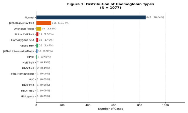
Figure 1. Distribution of haemoglobin patterns in 1077 paediatric samples by HPLC (N=1077).
Spectrum of Abnormal Haemoglobin Variants
β-Thalassemia trait was the most common abnormality, accounting for 116 cases (10.77% of total; 50.43% of abnormal samples). HPLC characteristically showed HbA₂ 3.5–8.0% with elevated HbA₂ peak and reduced HbA, correlating with microcytic hypochromic red cell indices (mean MCV 65.2 ± 7.4 fL, MCH 20.1 ± 3.8 pg). Unknown HPLC peaks were identified in 39 cases (3.62%), warranting further molecular work-up. Sickle cell trait was observed in 17 cases (1.58%), and homozygous sickle cell anaemia (HbSS) in 16 cases (1.49%). Details of abnormal variants are presented in Figure 2.
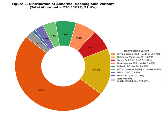
Figure 2. Proportional distribution of abnormal haemoglobin variants among 230 affected samples.
Haematological Parameters in Major Diagnostic Groups
Mean haematological and HPLC fraction data across the principal diagnostic categories are presented in Table 2 and Figure 3.
Table 2: Mean Haematological Indices and HPLC Fractions by Diagnostic Category (Mean ± SD)
|
Parameter |
Normal |
β-Thal Trait |
β-Thal Major |
HbAS Trait |
HbSS |
HPFH |
HbE Trait |
|
Hb (g/dL) |
11.8±1.6 |
9.6±1.8 |
6.2±1.4 |
11.2±1.5 |
7.4±1.9 |
11.0±1.4 |
10.1±1.2 |
|
MCV (fL) |
82.4±6.2 |
65.2±7.4 |
62.8±9.1 |
79.8±8.4 |
76.2±8.8 |
80.1±7.6 |
66.4±6.8 |
|
MCH (pg) |
27.6±3.2 |
20.1±3.8 |
18.4±4.1 |
25.4±3.6 |
24.2±3.4 |
26.4±3.2 |
19.8±3.1 |
|
HbA (%) |
96.14±1.4 |
84.40±3.8 |
19.10±12.4 |
57.60±8.6 |
9.52±4.8 |
64.20±8.2 |
56.40±9.2 |
|
HbA₂ (%) |
2.34±0.28 |
3.30±0.52 |
4.12±0.68 |
2.84±0.42 |
3.46±0.54 |
2.24±0.38 |
3.12±0.48 |
|
HbF (%) |
1.18±0.64 |
2.10±0.92 |
79.56±16.8 |
1.84±0.76 |
15.90±6.4 |
32.40±10.6 |
2.40±0.88 |
|
HbS (%) |
0 |
0 |
0 |
38.80±6.4 |
71.20±8.6 |
0 |
0 |
|
HbE (%) |
0 |
0 |
0 |
0 |
0 |
0 |
38.20±7.2 |
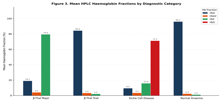
Figure 3. Grouped bar chart of mean HPLC fractions (HbA, HbA₂, HbF, HbS) across major diagnostic categories.
HPLC Patterns of Key Haemoglobin Variants
The characteristic HPLC retention-time patterns and diagnostic cut-offs for the main variants identified in this study are summarised in Table 3 and visualised as a diagnostic heatmap in Figure 4.
Peripheral Smear Correlation
Peripheral smear findings supported HPLC interpretation in the majority of cases. Microcytosis with target cells was consistently observed in β-thalassemia trait and HbE trait; marked anisopoikilocytosis with nucleated RBCs and polychromasia was characteristic of β-thalassemia major/intermedia (Table 4). Sickled erythrocytes with polychromasia and Howell–Jolly bodies were identified in all 16 cases of homozygous sickle cell anaemia; target cells and occasional sickled cells were seen in sickle cell trait.
Table 3: Characteristic HPLC Profiles of Key Haemoglobin Variants (Bio-Rad D10 System)
|
Condition |
HbA (%) |
HbA₂ (%) |
HbF (%) |
Variant Peak |
Diagnostic Clue |
|
Normal |
>95 |
1.5–3.5 |
<1 |
None |
Flat baseline |
|
β-Thal Trait |
83–95 |
3.5–8.0 |
0.5–3.5 |
None |
Elevated HbA₂ |
|
β-Thal Major |
5–30 |
3.5–6.0 |
60–98 |
None |
Massive HbF; absent/trace HbA |
|
HbAS (SCT) |
55–65 |
2.5–3.8 |
<2 |
HbS 30–45% |
HbS < HbA |
|
HbSS (SCA) |
<15 |
3.0–4.5 |
5–20 |
HbS 65–95% |
HbS >> HbA; positive sickling |
|
HPFH |
>75 |
1.5–2.8 |
15–35 |
None |
Raised HbF, normal indices |
|
HbE Trait |
70–80 |
2.8–3.6 |
<2 |
HbE 20–30% |
Peak at window 6 |
|
HbD-Punjab Trait |
70–80 |
2.5–3.4 |
<2 |
HbD 20–30% |
Co-elutes with HbS; negative sickling |
|
HbS+HbD |
5–15 |
3.5–4.5 |
5–12 |
HbS+HbD ≥75% |
Sickling positive; HbD co-migrates |
|
Hb Lepore |
75–85 |
2.0–2.8 |
<2 |
Lepore 5–15% |
P3 window; confirm by sequencing |
Sickling Test Results
Sodium metabisulphite sickling test was performed in 33 patients with HPLC peaks in the S-window. All 16 cases of homozygous sickle cell anaemia (HbSS) showed strongly positive sickling. Of 17 sickle cell trait cases, 15 (88.2%) were positive; 2 (11.8%) showed a delayed positive result at 120 minutes, likely reflecting lower concentrations of HbS. In one case with an S-window peak and negative sickling, further work-up revealed HbD-Punjab, confirming that co-elution of HbD with HbS on D10 requires the sickling test for definitive differentiation. The compound heterozygous HbS+HbD case showed a positive sickling test with a complex HPLC pattern.
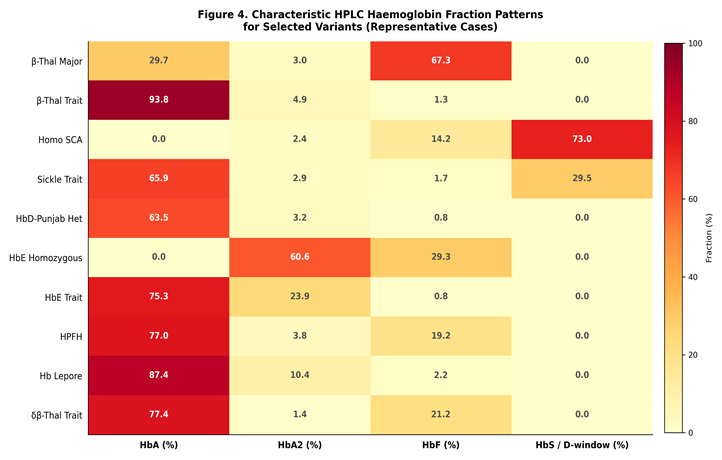
Figure 4. Diagnostic HPLC pattern heatmap showing characteristic haemoglobin fraction profiles for 10 variant types.
Table 4: Peripheral Blood Smear Morphology by Haemoglobin Condition
|
Condition |
Key Peripheral Smear Features |
|
β-Thalassemia Trait |
Mild microcytosis, hypochromia, occasional target cells, basophilic stippling; no nRBCs |
|
β-Thalassemia Major/Intermedia |
Severe anisopoikilocytosis, microcytosis, hypochromia, target cells, nRBCs (>5/100 WBCs), polychromasia, Howell–Jolly bodies |
|
Sickle Cell Trait (HbAS) |
Near-normal morphology; occasional target cells; no sickled cells under normal oxygenation |
|
Homozygous SCA (HbSS) |
Sickled erythrocytes (5–20%), target cells, polychromasia, nRBCs, Howell–Jolly bodies, thrombocytosis |
|
HPFH |
Normocytic/mildly microcytic; minimal morphological abnormality despite raised HbF |
|
HbE Trait |
Mild microcytosis, hypochromia, target cells; resembles β-thalassemia trait on smear |
|
HbD-Punjab Trait |
Essentially normal; no sickling despite positive S-window peak |
|
HbC Trait |
Target cells, occasional irregular contracted cells; mild microcytosis |
|
Hb Lepore |
Microcytic hypochromic anaemia with target cells; resembles β-thalassemia trait |
Molecular Findings
Molecular testing was performed in three children with severe transfusion-dependent anaemia showed homozygous c.92+5G>C (IVS-I-5 G>C) mutation in the HBB gene, confirming β-thalassemia major a splice-site mutation that leads to aberrant pre-mRNA processing and severely reduced β-globin production. This mutation is among the most common severe β-thalassemia mutations in South Asia.
Molecular results are summarised in Table 5.
Table 5: Summary of Molecular Analysis Findings
|
No. |
HPLC Finding |
Gene |
Mutation |
Diagnosis Confirmed |
|
1–3 |
Unknown peak; transfusion-dependent anaemia |
HBB |
c.92+5G>C (IVS-I-5 G>C) homozygous |
β-Thalassemia Major (3 children) |
Comparison with Published Indian Studies
The prevalence rates in the present study were compared with published Indian HPLC-based paediatric and general population studies. As depicted in Figure 5, the β-thalassemia trait prevalence (10.77%) in our series is consistent with values reported from comparable tertiary centres, though slightly lower than some high-prevalence community studies.
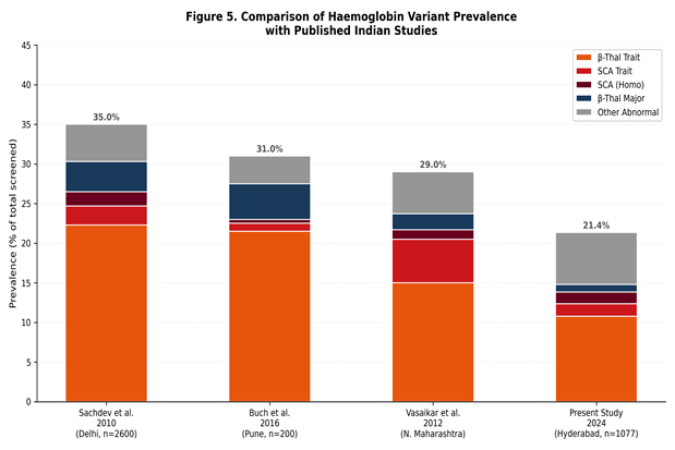
Figure 5. Comparative prevalence of major haemoglobin variants (β-thalassemia trait and sickle cell trait) across published Indian HPLC studies.
DISCUSSION:
The present study documents the spectrum of hemoglobinopathies in a large paediatric cohort (n=1077) at a tertiary care centre in Telangana using a comprehensive diagnostic workflow. The overall prevalence of abnormal haemoglobin patterns was 21.35%, highlighting the significant burden of these disorders in the catchment population. This figure is broadly concordant with estimates from comparable Indian tertiary-care studies, which report abnormal HPLC patterns in 15–35% of paediatric samples.1,2,15
β-Thalassemia Trait
β-Thalassemia trait was the predominant abnormality (116 cases, 10.77%), consistent with its reported prevalence of 3–17% in various Indian populations.5,6 Osmania Medical College and Niloufer Hospital serve Hyderabad’s multiethnic population, which includes communities such as Muslims, Brahmins, and certain tribal groups known to have elevated carrier frequencies due to consanguineous marriages and endogamy.7 The HPLC hallmark of β-thalassemia trait is HbA₂ >3.5% with concurrent microcytosis (MCV <80 fL) and hypochromia (MCH <27 pg).12 One diagnostic challenge is the co-existence of iron-deficiency anaemia (IDA) with β-thalassemia trait, which can reduce HbA₂ into the borderline range (3.2–3.5%), leading to under-diagnosis.13 In our study, iron studies were not uniformly available; this limitation may have resulted in a few missed borderline diagnoses. Concurrent iron therapy and ferritin estimation is therefore recommended before HPLC-based screening.
Sickle Cell Disorders
Sickle cell trait (HbAS) was identified in 17 cases (1.58%) and homozygous sickle cell anaemia (HbSS) in 16 cases (1.49%). While these rates may appear low compared to Vidarbha or Chhattisgarh regions with >20% HbS carrier frequency Hyderabad’s mixed urban population yields intermediate values.16,17 The sodium metabisulphite sickling test remained a cost-effective and reliable adjunct: all 16 HbSS cases were strongly positive, and 15 of 17 trait cases tested positive. Notably, in one case with an HPLC peak in the S-window and a negative sickling test, HbD-Punjab was confirmed by molecular analysis. This underscores the fundamental principle that the sickling test is mandatory whenever an S-window peak is detected on HPLC, as HbD-Punjab co-elutes with HbS on the D10 system at identical retention times.18
The compound heterozygous HbS+HbD-Punjab (1 case) presented with a severe sickle cell phenotype similar to HbSS disease, characterised by positive sickling, splenomegaly, and haemolytic anaemia. Published literature confirms that HbS/HbD-Punjab compound heterozygotes may exhibit clinical severity comparable to HbSS, partly because HbD-Punjab does not inhibit HbS polymerisation.19
β-Thalassemia Major/Intermedia
β-Thalassemia major and intermedia accounted for 10 cases (0.93%). Molecularly, three of these children were confirmed to carry the homozygous c.92+5G>C (IVS-I-5 G>C) mutation in the HBB gene. This splice-site mutation disrupts normal pre-mRNA splicing, activating a cryptic splice site within intron 1, leading to reduced (or absent) functional β-globin chain production and consequent severe transfusion-dependent anaemia.20 The IVS-I-5 G>C mutation is one of the four most prevalent severe β-thalassemia mutations in South Asia (alongside IVS-I-1 G>A, codon 8/9 +G, and codon 41/42 −TTCT), accounting for approximately 15–18% of Indian β-thalassemia alleles.20,21
HPLC in β-thalassemia major characteristically shows a very high HbF fraction (>60%), near-absent HbA, and mildly elevated HbA₂. The peripheral smear revealed severe anisopoikilocytosis, polychromasia, nucleated RBCs, and tear-drop cells. All three confirmed cases were transfusion-naïve at initial presentation and were subsequently enrolled in a regular transfusion and chelation programme.
HPFH and δβ-Thalassemia
Seven cases (0.65%) showed raised HbF with otherwise normal haematological parameters, consistent with hereditary persistence of fetal haemoglobin (HPFH). An additional 16 cases labelled "raised HbF/HbF variant" included both HPFH and δβ-thalassemia carriers. Distinguishing HPFH from δβ-thalassemia has clinical relevance: HPFH is benign, while δβ-thalassemia in compound heterozygosity with β-thalassemia or HbS may cause moderate disease. HPLC alone cannot reliably distinguish these entities; gap-PCR or MLPA for delta-beta globin gene deletions is recommended.22
Rare Variants: HbE, HbD, HbQ, HbC, and Hb Lepore
Rare variants—including HbE trait (2 cases), HbD-Punjab trait (2 cases), HbQ trait (1 case), HbC (1 case), and Hb Lepore (1 case)—collectively constituted 7 cases (3.04% of abnormal). These findings demonstrate the value of a high-sensitivity HPLC-based screening programme in a diverse population. HbE is the most prevalent haemoglobin variant globally after HbS and is highly prevalent in north-east India and South-East Asia.23 HbE in compound heterozygosity with β-thalassemia (βE-thalassemia) is the most common form of severe thalassaemia in South and South-East Asia, reinforcing the importance of HbE carrier detection.
HbQ-alpha is a rare structural variant (alpha-globin chain substitution) that elutes in the A2 window on D10 chromatograms and may raise the apparent HbA₂. Failure to recognise HbQ may falsely diagnose β-thalassemia trait. In our case, peripheral smear showed essentially normal morphology, and the "elevated HbA₂" was on review recognised as a distinct HbQ peak.24
Hb Lepore (one case) presented as a P3-window peak with mild thalassaemic indices. Hb Lepore arises from non-homologous crossover between the δ- and β-globin genes, producing a δβ-fusion chain. It is clinically and haematologically similar to β-thalassemia trait in the heterozygous state and may cause severe disease when homozygous or compound heterozygous with β-thalassemia. Confirmation by DNA sequencing was essential in this case.25
Unknown Peaks
Unknown peaks (39 cases, 3.62%) represented a significant diagnostic challenge. These included samples with variant peaks at unusual retention times not identifiable by the D10 software. Cases were referred to higher centres for advanced characterisation. This highlights a recognised limitation of HPLC: uncommon or novel variants generating atypical retention-time patterns require supplementary techniques, including DNA sequencing, for definitive diagnosis.14,26
Role of HPLC in Paediatric Screening
HPLC remains the most widely recommended first-line tool for neonatal and paediatric hemoglobinopathy screening globally and in Indian guidelines.27,28 Its advantages include: simultaneous quantification of HbA, HbA₂, HbF, and structural variants in a single run; high throughput (~200 samples/8-hour shift on D10); excellent reproducibility (CV <1% for major fractions); and the ability to detect rare variants. In resource-constrained settings, HPLC combined with a targeted peripheral smear and sickling test provides a highly cost-effective diagnostic algorithm.
The high prevalence of β-thalassemia trait (10.77%) and sickle cell trait (1.58%) in our paediatric cohort has direct public health implications. Universal neonatal screening would allow early prophylactic penicillin and pneumococcal vaccine administration in SCA patients, significantly reducing infectious morbidity; early identification of β-thalassemia major enables timely enrolment in transfusion and chelation programmes; and carrier detection enables genetic counselling before reproduction.29,30
Strengths and Limitations
Strengths of this study include the large paediatric sample size (n=1077), the comprehensive multimodal diagnostic workflow (CBC, peripheral smear, sickling test, HPLC, and selective molecular analysis), and the identification of rare variants including HbQ, Hb Lepore, HbS+HbD compound heterozygotes, and the c.92+5G>C β-thalassemia mutation.
Limitations include: (i) the retrospective design with incomplete clinical follow-up data; (ii) non-uniform availability of iron studies, which may have caused underestimation of β-thalassemia trait in iron-deficient subjects; (iii) single-centre data that may not reflect the full epidemiological diversity of the region; and (iv) molecular analysis was performed in only a subset of cases, leaving some unknown peaks uncharacterised.
CONCLUSION:
β-Thalassemia trait is the most common hemoglobinopathy in the paediatric population served by this tertiary centre, followed by sickle cell-related disorders and raised HbF variants. HPLC using the Bio-Rad D10 system is a sensitive and reliable first-line tool capable of identifying both common and rare haemoglobin variants, including HbQ, Hb Lepore, and HbS+HbD compound heterozygotes. Peripheral smear examination and the sodium metabisulphite sickling test remain indispensable adjuncts for resolving HPLC ambiguities. Molecular analysis is essential for unknown peaks, borderline cases, and confirmation of severe mutations such as c.92+5G>C in the HBB gene. The significant carrier burden identified in this study underscores the urgent need for structured neonatal and antenatal screening programmes, systematic genetic counselling, and comprehensive family studies to curtail the incidence of severe hemoglobinopathies in future generations.
REFERENCES:
1. Shankar R, Prasad KN, Banerjee BC. Role of HPLC as a tool for detection of hemoglobinopathies. IOSR J Dent Med Sci. 2019;18(8):1–5.
2. Buch A, Iqbal B, Bordawekar R, Jain A, Jariwala P, Rathod H. Patterns of hemoglobinopathies diagnosed by HPLC in Western Maharashtra. J Med Soc. 2016;30(2):111–5.
3. Modell B, Darlison M. Global epidemiology of haemoglobin disorders and derived service indicators. Bull World Health Organ. 2008;86(6):480–7.
4. Weatherall DJ. The inherited diseases of hemoglobin are an emerging global health burden. Blood. 2010;115(22):4331–6.
5. Balgir RS. Epidemiology, population health genetics and phenotypic diversity of sickle cell disease in India. Internet J Biol Anthropol. 2007;1(2).
6. Colah R, Gorakshakar A, Nadkarni A. Global burden, distribution and prevention of beta-thalassemias and hemoglobin E disorders. Expert Rev Hematol. 2010;3(1):103–17.
7. Weatherall DJ, Clegg JB. The Thalassemia Syndromes. 4th ed. Oxford: Blackwell Science; 2001.
8. Kumar V, Abbas AK, Aster JC. Robbins and Cotran Pathologic Basis of Disease. 9th ed. Philadelphia: Elsevier; 2015. p. 634–641.
9. Steinberg MH, Forget BG, Higgs DR, Weatherall DJ, eds. Disorders of Hemoglobin: Genetics, Pathophysiology, and Clinical Management. 2nd ed. Cambridge: Cambridge University Press; 2009.
10. Cao A, Galanello R. Beta-thalassemia. Genet Med. 2010;12(2):61–76.
11. Harteveld CL, Higgs DR. Alpha-thalassaemia. Orphanet J Rare Dis. 2010;5:13.
12. Joutovsky A, Hadzi-Nesic J, Nardi MA. HPLC retention time as a diagnostic tool for hemoglobin variants and hemoglobinopathies: a study of 60,000 samples in a clinical diagnostic laboratory. Clin Chem. 2004;50(10):1736–47.
13. Mosca A, Paleari R, Ivaldi G, Galanello R, Giordano PC. The role of haemoglobin A(2) in the diagnosis of beta-thalassemia trait. Haematologica. 2009;94(8):1143–48.
14. Wild BJ, Bain BJ. Investigation of abnormal haemoglobins and thalassaemia. In: Dacie and Lewis Practical Haematology. 11th ed. Churchill Livingstone; 2011. p. 271–314.
15. Sachdev R, Dam AR, Tyagi G. Detection of Hb variants and hemoglobinopathies in Indian population using HPLC: report of 2600 cases. Indian J Pathol Microbiol. 2010;53(1):57–62.
16. Mohanty D, Mukherjee MB, Colah RB. Sickle cell disease in India. Curr Opin Hematol. 2004;11(3):178–82.
17. Yanni E, Grosse SD, Yang Q, Olney RS. Trends in pediatric sickle cell disease-related mortality in the United States, 1983–2002. J Pediatr. 2009;154(4):541–5.
18. Clarke GM, Higgins TN. Laboratory investigation of hemoglobinopathies and thalassemias: review and update. Clin Chem. 2000;46(8 Pt 2):1284–90.
19. Serjeant GR, Serjeant BE, Boucher-Hayes D, Hambleton IR. Haemoglobin S/D (Punjab) disease: a clinical and haematological description. J Trop Med Hyg. 1993;96(2):105–11.
20. Thein SL. Molecular basis of beta thalassemia and potential therapeutic targets. Blood Cells Mol Dis. 2018;70:54–65.
21. Colah R, Gorakshakar A, Phanasgaonkar S, D’Souza E, Nadkarni A, Surve R, et al. Epidemiology of beta-thalassaemia in Maharashtra, India: mapping the frequencies and mutations in sub-regions of Maharashtra. Eur J Haematol. 2010;84(1):45–52.
22. Galanello R, Barella S, Turco MP, Giagheddu M, Perseu L, Ximenes A, et al. Serum erythropoietin and erythropoiesis in high- and low-fetal hemoglobin beta-thalassemia intermedia patients. Blood. 1994;83(2):561–5.
23. Fucharoen S, Winichagoon P. Haemoglobinopathies in Southeast Asia. Indian J Med Res. 2011;134(4):498–506.
24. Adekile AD, Haider MZ, Chibber S. Hb Q-India in Kuwait with mild clinical picture. Hemoglobin. 2003;27(2):89–94.
25. Olivieri NF, Weatherall DJ. The hemoglobinopathies. In: Harrison’s Principles of Internal Medicine. 19th ed. McGraw-Hill; 2015. p. 631–641.
26. Paleari R, Mosca A, Zoppi F, Brambilla S, Girelli G. Haemoglobin reference intervals for an Italian population: update using the new Bio-Rad Variant™ II system. Scand J Clin Lab Invest. 2009;69(5):525–32.
27. Gibbons R. Hemoglobinopathy — WHO perspectives on prevention and management. WHO Tech Rep Ser. 2006;938:1–80.
28. Indian Council of Medical Research. Guidelines for the management of thalassaemias and sickle cell disease. ICMR Task Force Study. New Delhi: ICMR; 2020.
29. Wambua S, Nkya S, Mwakuziwe S, Makani J, Kirungi A, Mwangi TW, et al. WHO sickle-cell disease burden; the management of sickle-cell disease in sub-Saharan Africa. Lancet Haematol. 2020;7(8):e585–e591.
30. Shanthala DM, Sitalakshmi S, Parameshwar RD, Prasad EV. Study of hemoglobinopathies and thalassemias using HPLC (NBS method) in a tertiary care hospital. Indian J Hematol Blood Transfus. 2008;24(2):72–6.