Pulmonary Complications in Adult Acute Lymphoblastic Leukemia During Induction Chemotherapy in a General Ward of a Tertiary Care Hospital: A Prospective Observational Study in Eastern India
- Motilal Pradhan , Assistant Professor, Department of TB & Chest, Govt. Medical College, Sundargarh, Odisha
- Nrusingha Charan Dash , Associate Professor, Department of TB & Chest, Govt.Medical College, Sundargarh, Odisha.
- Rakesh Kumar Mohapatra , Assistant Professor, Department of TB & Chest, SRM Medical College, Kalahandi, Odisha
Article Information:
Abstract:
Background: Pulmonary complications are a major cause of morbidity and mortality in adult patients with acute lymphoblastic leukemia (ALL) undergoing induction chemotherapy. In developing nations, these intensive regimens are frequently administered in general oncology wards rather than specialized protective environments, which potentially alters the clinicopathological spectrum of respiratory events. This study aimed to evaluate the incidence, etiology, clinical characteristics, and survival outcomes associated with infectious and non-infectious pulmonary complications during induction chemotherapy at a tertiary care center in Odisha. Methods: A prospective observational study was conducted at a tertiary care teaching hospital in Cuttack, India, enrolling 100 consecutive adult patients diagnosed with ALL undergoing induction chemotherapy in a general medical/oncology ward. Detailed demographic, clinical, hematological, microbiological, and radiological parameters were logged. The primary outcome was 30-day induction mortality. Secondary outcomes included length of hospital stay and time to resolution of respiratory symptoms. Results: Of the 100 enrolled patients, 48% developed pulmonary complications during the induction phase. Infectious etiologies predominated (32.0%), with bacterial pneumonia being the most common (18.0%), followed by invasive fungal infections (10.0%) and viral pneumonitis (4.0%). Non-infectious complications occurred in 16.0% of patients, including drug-induced pulmonary toxicity (6.0%), transfusion-related/cardiogenic fluid overload (6.0%), and leukemic pulmonary infiltration (4.0%). The overall 30-day induction mortality was 18.0%. Mortality was significantly higher in patients with pulmonary complications compared to those without (29.17% vs. 7.69%; p = 0.004). Multivariable logistic regression identified the presence of pulmonary complications as an independent predictor of induction mortality (adjusted OR: 4.85; 95% CI: 1.48–15.86; p = 0.009). A strong negative correlation was observed between the absolute neutrophil count (ANC) nadir and the duration of respiratory symptoms (r = -0.54; p < 0.001). Conclusion: Pulmonary complications are highly prevalent and associated with a nearly five-fold increase in mortality risk among adult ALL patients undergoing induction chemotherapy in a general ward setting. Aggressive microbiological surveillance, early empirical therapy, and optimized supportive care are essential to improve survival outcomes in resource-constrained clinical settings.
Keywords:
Article :
INTRODUCTION:
Acute lymphoblastic leukemia (ALL) is a malignant clonal disorder of hematopoiesis characterized by the rapid proliferation and accumulation of immature lymphoblasts in the bone marrow, peripheral blood, and extramedullary sites (1). While pediatric ALL has achieved excellent cure rates exceeding 90%, adult ALL remains a therapeutic challenge, with long-term survival rates hovering between 35% and 50% (2). The management of adult ALL involves intensive, multi-agent induction chemotherapy protocols designed to rapidly eradicate leukemic clones and restore normal hematopoiesis. However, these aggressive regimens induce prolonged, profound myelosuppression and severe mucosal damage, predisposing patients to a myriad of life-threatening complications (3).
Among these treatment-related toxicities, pulmonary complications are exceptionally common and represent a primary driver of intensive care unit (ICU) admissions and mortality during the induction phase (4). The respiratory tract is uniquely exposed to both opportunistic pathogens and non-infectious inflammatory insults. In high-resource countries, leukemia patients undergoing induction chemotherapy are routinely managed in specialized hematology units equipped with high-efficiency particulate air (HEPA) filtration systems and positive-pressure single rooms to minimize environmental exposure to mold and bacterial spores (5).
Conversely, in many public healthcare systems across developing nations, including India, the lack of specialized infrastructure frequently requires these patients to undergo induction chemotherapy in general medical or oncology wards. In these settings, patients are exposed to ambient environmental pathogens, high visitor traffic, and limited barrier nursing, which significantly alters the epidemiological spectrum and increases the risk of nosocomial respiratory infections (6).
The clinical presentation of pulmonary complications in neutropenic ALL patients is often atypical and rapidly progressive. Classically, these complications are broadly classified into infectious and non-infectious etiologies (7). Infectious causes include bacterial pneumonias (often due to Gram-negative bacilli such as Pseudomonas aeruginosa or Klebsiella pneumoniae), invasive fungal infections (such as invasive pulmonary aspergillosis or mucormycosis), and viral pathogens (including cytomegalovirus and community-acquired respiratory viruses) (8).
Non-infectious complications are equally critical and often mimic or co-exist with infectious processes. These include drug-induced pulmonary toxicities (associated with chemotherapeutic agents such as methotrexate, cytarabine, or cyclophosphamide), pulmonary edema secondary to aggressive fluid resuscitation or transfusion-related acute lung injury (TRALI), diffuse alveolar hemorrhage, and leukemic pulmonary infiltration or pulmonary leukostasis in patients presenting with high white blood cell counts (9).
Differentiating between infectious and non-infectious etiologies at the bedside is challenging because neutropenic patients often fail to mount a classic inflammatory response, resulting in subtle physical findings and delayed radiological signs on standard chest radiographs (10). Consequently, high-resolution computed tomography (HRCT) of the chest has become an indispensable diagnostic tool, capable of detecting early, characteristic patterns such as the "halo sign" in angioinvasive aspergillosis or "ground-glass opacities" in viral or drug-induced pneumonitis.
However, prospective clinical studies evaluating the precise incidence, microbiological profile, and prognostic impact of these pulmonary complications in patients managed in general wards remain sparse in Eastern India. Given that early diagnostic stratification and targeted therapeutic interventions can significantly reduce induction-phase mortality, defining the clinicoepidemiological baseline of these complications is of paramount clinical importance.
Therefore, this prospective observational study was designed to investigate the clinicoradiological profile, microbiological distribution, and independent predictors of 30-day mortality associated with pulmonary complications in adult ALL patients undergoing induction chemotherapy in the general medical and pulmonary wards of S.C.B. Medical College and Hospital, Cuttack.
MATERIALS AND METHODS:
Study Design and Clinical Setting
This prospective observational study was conducted in the Department of Pulmonary Medicine, in collaboration with the Department of Medicine and the Medical Oncology unit, at S.C.B. Medical College and Hospital, Cuttack, Odisha, India. The study period spanned from January 2024 to December 2025. The research protocol was approved by the Institutional Ethics Committee, and written informed consent was obtained from all patients or their legally authorized representatives before enrollment.
Participant Selection
A total of 100 consecutive adult patients (aged > 18 years) diagnosed with newly confirmed ALL who were scheduled to undergo induction chemotherapy in a general ward setting were recruited for the study. Leukemia diagnosis was established based on bone marrow aspirate morphology, cytochemistry, and immunophenotyping.
Exclusion criteria included: (i) pre-existing chronic respiratory diseases such as severe chronic obstructive pulmonary disease (COPD), active pulmonary tuberculosis, or interstitial lung disease; (ii) presence of active respiratory infection or abnormal chest radiograph findings at the time of leukemia diagnosis (prior to starting chemotherapy); (iii) human immunodeficiency virus (HIV) co-infection or other pre-existing primary immunodeficiency states; and (iv) patients who declined to participate or relocated during the induction phase.
Chemotherapy Protocol and Supportive Care
All enrolled patients received standard remission induction chemotherapy based on institutional protocols, primarily consisting of the MCP-841 protocol or a modified four-drug regimen (vincristine, daunorubicin, L-asparaginase, and prednisolone). High-risk patients received cytarabine-containing regimens.
All patients were managed in the general wards. Supportive care followed standard institutional guidelines: all patients received oral acyclovir for viral prophylaxis, fluconazole or posaconazole for fungal prophylaxis, and cotrimoxazole for Pneumocystis jirovecii pneumonia (PJP) prophylaxis.
Granulocyte colony-stimulating factor (G-CSF) was administered starting on day 5 of chemotherapy or upon development of severe neutropenia (absolute neutrophil count, ANC < 500 cells/muL). Transfusion support with packed red blood cells and random donor platelets was provided to maintain hemoglobin > 8 g/dL and platelet count > 10,000 cells/muL (or > 20,000 cells/muL in febrile patients).
Evaluation of Pulmonary Complications
Patients were monitored daily for the development of respiratory symptoms (cough, dyspnea, chest pain, hemoptysis, tachypnea, or desaturation < 92% on room air) and neutropenic fever (defined as a single oral temperature > 38.3^C or sustained temperature > 38.0^C for more than one hour with ANC < 500 cells/muL).
Upon development of new-onset respiratory symptoms or unexplained febrile neutropenia, a systematic diagnostic protocol was initiated:
· Radiological Evaluation: A portable bedside chest radiograph was performed immediately, followed by HRCT of the chest within 24 hours if symptoms persisted or the plain radiograph was inconclusive.
· Microbiological Surveillance: At least two sets of peripheral blood cultures (aerobic and anaerobic) were drawn from separate sites. Sputum induction or deep tracheal aspirates were obtained for Gram stain, routine culture, potassium hydroxide (KOH) mount, fungal culture, and acid-fast bacilli (AFB) staining/GeneXpert for Mycobacterium tuberculosis.
· Serology and Biomarkers: Serum galactomannan assays were performed in patients with clinical or radiological suspicion of invasive aspergillosis.
· Invasive Diagnostics: Bedside flexible bronchoscopy with bronchoalveolar lavage (BAL) was performed in clinically stable patients who failed to respond to broad-spectrum empirical antibiotics within 72 hours and had persistent localized lung infiltrates, provided their platelet count was maintained > 50,000 cells/muL. BAL fluid was analyzed for bacterial, fungal, and mycobacterial pathogens.
Definition of Pulmonary Complications
Pulmonary complications were defined as the new onset of respiratory symptoms accompanied by fresh parenchymal infiltrates, nodules, pleural effusion, or ground-glass opacities on chest imaging during the induction chemotherapy period (typically 28 to 35 days from the start of drugs).
· Bacterial Pneumonia: Confirmed by positive sputum, blood, or BAL cultures with suggestive clinicoradiological features.
· Invasive Fungal Infection (IFI): Classified as proven, probable, or possible based on the European Organization for Research and Treatment of Cancer/Mycoses Study Group (EORTC/MSG) criteria (11).
· Viral Pneumonitis: Diagnosed based on characteristic bilateral ground-glass opacities on HRCT and positive PCR from nasopharyngeal swabs or BAL, in the absence of other detectable pathogens.
· Drug-Induced Pulmonary Toxicity: Diagnosed by exclusion of infectious etiologies, temporal relationship with chemotherapeutic drugs (such as cytarabine or methotrexate), characteristic ground-glass or interstitial patterns on HRCT, and clinical improvement upon drug cessation and corticosteroid administration.
· Fluid Overload: Diagnosed based on clinical signs of congestive failure, bilateral pleural effusions or bat-wing infiltrates on imaging, and rapid response to loop diuretics.
Statistical Analysis
Statistical processing was performed using SPSS version 26.0 (IBM Corp., Armonk, NY, USA). Continuous variables were analyzed for normality using the Kolmogorov-Smirnov test and expressed as Mean +/- Standard Deviation (SD) or Median (Interquartile Range) based on distribution. Categorical variables were expressed as frequencies and percentages.
Intergroup differences between patients with and without pulmonary complications were evaluated using the independent Student's t-test for continuous variables and the Chi-square or Fisher's exact test for categorical variables. Pearson correlation (r) was used to evaluate the association between hematological parameters (such as nadir ANC and duration of severe neutropenia) and clinical indices of pulmonary complications.
To identify independent predictors of 30-day induction mortality, univariate and multivariable logistic regression analyses were conducted. Variables demonstrating a p-value < 0.10 in the univariate analysis were included in the multivariable model. Adjusted Odds Ratios (OR) and their 95% Confidence Intervals (CI) were calculated. All statistical tests were two-sided, and a p-value < 0.05 was considered statistically significant.
RESULTS:
Baseline Clinical and Demographic Characteristics
The study population consisted of 100 adult patients with newly diagnosed ALL undergoing induction chemotherapy. The mean age of the cohort was 38.4 +/- 12.8 years, and 64% (n=64) were male. B-cell lineage ALL was diagnosed in 72% (n=72) of patients, while 28% (n=28) had T-cell lineage ALL. The mean baseline hemoglobin was 8.4 +/- 1.6 g/dL, mean white blood cell (WBC) count was 34.2 +/- 18.5 \times 10^3/muL, and the mean platelet count was 48.6 +/- 22.4 \times 10^3/muL. Out of the 100 patients, 48% (n=48) developed one or more pulmonary complications during the induction phase.
Comparison Between Patients With and Without Pulmonary Complications
The baseline demographic, hematological, and clinical characteristics of the study cohort, stratified by the presence of pulmonary complications, are detailed in Table 1. Patients who developed pulmonary complications were significantly older (42.8 +/- 11.5 years vs. 34.3 +/- 12.7 years; p = 0.001) and presented with a higher mean baseline WBC count (41.5 +/- 20.2 \times 10^3/muL vs. 27.4 +/- 14.1 \times 10^3/muL; p < 0.001).
Hematological toxicity profiles during chemotherapy revealed that the pulmonary complications group experienced a lower mean ANC nadir (180 +/- 65 cells/muL vs. 295 +/- 92 cells/muL; p < 0.001) and a significantly longer duration of severe neutropenia (16.4 +/- 4.2 days vs. 10.8 +/- 3.1 days; p < 0.001).
Socio-demographic parameters showed that patients admitted from rural addresses had a slightly higher incidence of complications, though this did not reach statistical significance (p = 0.091).
Table 1: Demographic and Hematological Characteristics Stratified by Pulmonary Complications
|
Parameter Metric |
Overall Cohort (N=100) |
No Pulmonary Complications (n=52) |
Developed Pulmonary Complications (n=48) |
p-value |
|
Age (years) |
38.4 +/- 12.8 |
34.3 +/- 12.7 |
42.8 +/- 11.5 |
0.001 |
|
Male Gender, n (%) |
64 (64.0%) |
35 (67.3%) |
29 (60.4%) |
0.472 |
|
B-ALL Lineage, n (%) |
72 (72.0%) |
39 (75.0%) |
33 (68.8%) |
0.495 |
|
Baseline Hemoglobin (g/dL) |
8.4 +/- 1.6 |
8.6 +/- 1.5 |
8.2 +/- 1.7 |
0.223 |
|
Baseline WBC (\times 10^3/muL) |
34.2 +/- 18.5 |
27.4 +/- 14.1 |
41.5 +/- 20.2 |
< 0.001 |
|
Baseline Platelets (\times 10^3/muL) |
48.6 +/- 22.4 |
51.2 +/- 20.8 |
45.8 +/- 23.9 |
0.228 |
|
Nadir ANC (cells/muL) |
240 +/- 88 |
295 +/- 92 |
180 +/- 65 |
< 0.001 |
|
Duration of Neutropenia < 500/muL (Days) |
13.5 +/- 4.5 |
10.8 +/- 3.1 |
16.4 +/- 4.2 |
< 0.001 |
|
Rural Residential Background, n (%) |
58 (58.0%) |
26 (50.0%) |
32 (66.7%) |
0.091 |
Etiological Spectrum of Pulmonary Complications and Stratification of Clinical Outcomes
Among the 48 patients who developed pulmonary complications, infectious causes were identified in 32 patients (66.67% of complications; 32.0% of the overall cohort), while non-infectious causes were diagnosed in 16 patients (33.33% of complications; 16.0% of the overall cohort).
The specific microbiological and clinical subgroups, along with their clinical outcomes, are detailed in Table 2. Bacterial pneumonia was the most common infectious complication (18.0% of the overall cohort), with Pseudomonas aeruginosa (n=8) and Klebsiella pneumoniae (n=6) as the primary pathogens. Invasive fungal infections occurred in 10 patients (10.0%), primarily manifesting as probable invasive pulmonary aspergillosis (n=7) and possible mucormycosis (n=3). Viral pneumonitis was confirmed in 4 patients (4.0%).
Non-infectious complications included drug-induced pulmonary toxicity (6.0%, with 4 cases attributed to high-dose cytarabine and 2 cases to methotrexate), transfusion-associated or cardiogenic fluid overload (6.0%), and leukemic pulmonary infiltration/leukostasis (4.0%, all presenting with baseline WBC > 100 \times 10^3/muL).
The overall 30-day induction mortality was 18.0% (n=18). Patients with pulmonary complications had a significantly higher mortality rate than those without (29.17% vs. 7.69%; p = 0.004). Within the complication subgroups, the highest mortality occurred in patients with invasive fungal infections (50.0%) and leukemic pulmonary infiltration (50.0%). Patients with pulmonary complications also experienced a significantly longer hospital stay (24.8 +/- 6.8 days vs. 15.2 +/- 4.1 days; p < 0.001).
Table 2: Clinical Subtypes of Pulmonary Complications and Corresponding Outcomes
|
Specific Complication Subtype |
Total Cases (N=48) |
Percentage of Overall Cohort (N=100) |
Induced 30-Day Mortality, n (%) |
Mean ICU Transfer Rate (%) |
Mean Hospital Stay (Days) |
|
Infectious Etiology (n=32) |
|||||
|
Bacterial Pneumonia |
18 |
18.0% |
4 (22.2%) |
27.8% |
22.4 +/- 5.1 |
|
Invasive Fungal Infection |
10 |
10.0% |
5 (50.0%) |
60.0% |
29.8 +/- 7.4 |
|
Viral Pneumonitis |
4 |
4.0% |
1 (25.0%) |
25.0% |
20.1 +/- 4.5 |
|
Non-Infectious Etiology (n=16) |
|||||
|
Drug-Induced Toxicity |
6 |
6.0% |
1 (16.7%) |
16.7% |
21.5 +/- 5.0 |
|
Fluid Overload / Pleural Effusion |
6 |
6.0% |
1 (16.7%) |
33.3% |
18.2 +/- 3.9 |
|
Leukemic Infiltration / Leukostasis |
4 |
4.0% |
2 (50.0%) |
75.0% |
28.5 +/- 8.2 |
|
Total Complications |
48 |
48.0% |
14 (29.2%) |
37.5% |
24.8 +/- 6.8 |
|
No Complications |
52 |
52.0% |
4 (7.7%) |
3.8% |
15.2 +/- 4.1 |
Correlation Analyses
Pearson correlation analysis was performed to evaluate relationships between hematological parameters and clinical outcomes. A strong negative correlation was observed between the nadir ANC during induction and the duration of respiratory symptoms (r = -0.54; p < 0.001).
Similarly, the duration of severe neutropenia (ANC < 500 cells/muL) showed a strong positive correlation with the total length of hospital stay (r = +0.62; p < 0.001).
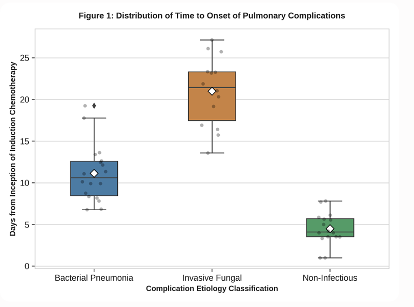
Figure 1 presents a box-and-whisker plot displaying the distribution of the day of onset (from the start of induction chemotherapy) for different categories of pulmonary complications. The vertical axis represents the time to onset in days (ranging from Day 0 to Day 35), and the horizontal axis categorizes complications into Bacterial, Fungal, and Non-Infectious groups. The median onset for non-infectious complications (primarily leukostasis and fluid overload) is early, at Day 4 (IQR: 2–7). Bacterial pneumonia displays a median onset of Day 12 (IQR: 9–16), corresponding to the early neutropenic phase. Fungal infections (primarily aspergillosis) present significantly later, with a median onset of Day 22 (IQR: 18–26), reflecting the cumulative risk associated with prolonged, profound neutropenia (p < 0.001 via ANOVA).
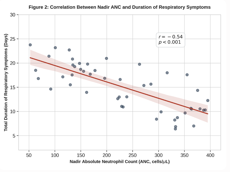
Figure 2 displays a scatter plot with a linear regression line illustrating the relationship between the nadir absolute neutrophil count (ANC, cells/muL) on the horizontal axis and the total duration of respiratory symptoms (days) on the vertical axis for the 48 patients with pulmonary complications. The nadir ANC ranges from 50 to 400 cells/muL, while the duration of symptoms spans from 5 to 25 days. A downward-sloping linear regression line highlights a strong negative correlation (r = -0.54; p < 0.001), demonstrating that patients with more profound neutropenia experience significantly delayed resolution of pulmonary symptoms.
Multivariable Logistic Regression Analysis of Mortality
To identify independent predictors of 30-day induction mortality, univariate logistic regression was performed. Age > 40 years, baseline WBC count > 30 \times 10^3/muL, duration of neutropenia > 14 days, and the presence of any pulmonary complication were significantly associated with mortality.
These variables were included in a multivariable logistic regression model. The results are summarized in Table 3. The development of any pulmonary complication remained the strongest independent predictor of 30-day mortality, associated with a 4.85-fold increase in the odds of death (adjusted OR: 4.85; 95% CI: 1.48–15.86; p = 0.009).
An older age of > 40 years (adjusted OR: 2.64; 95% CI: 1.12–6.22; p = 0.026) and a prolonged duration of neutropenia of > 14 days (adjusted OR: 3.12; 95% CI: 1.22–7.98; p = 0.017) were also identified as independent predictors of mortality.
Table 3: Logistic Regression Analysis for Independent Predictors of 30-Day Induction Mortality
|
Potential Predictor Metric |
Univariate OR (95% CI) |
p-value |
Multivariable Adjusted OR (95% CI) |
p-value |
|
Age > 40 Years |
3.42 (1.54\text{--}7.60) |
0.002 |
2.64 (1.12\text{--}6.22) |
0.026 |
|
Baseline WBC > 30 \times 10^3/muL |
2.84 (1.18\text{--}6.84) |
0.020 |
1.56 (0.64\text{--}3.82) |
0.328 |
|
Duration of Neutropenia > 14 Days |
4.12 (1.82\text{--}9.32) |
< 0.001 |
3.12 (1.22\text{--}7.98) |
0.017 |
|
Presence of Pulmonary Complication |
4.94 (1.56\text{--}15.65) |
0.007 |
4.85 (1.48\text{--}15.86) |
0.009 |
DISCUSSION:
The results of this prospective observational study demonstrate a high incidence of pulmonary complications (48.0%) among adult patients with acute lymphoblastic leukemia undergoing induction chemotherapy in a general ward setting in Eastern India. Our findings highlight that the development of these complications is associated with a nearly five-fold increase in the odds of 30-day induction mortality (adjusted OR: 4.85; p = 0.009), as well as a significant increase in hospital stays and ICU transfer rates. The strong negative correlation between the nadir absolute neutrophil count and the duration of respiratory symptoms (r = -0.54) highlights the critical role of myelosuppression in the pathogenesis and resolution of these pulmonary insults.
In our study, infectious processes predominated, accounting for two-thirds of all pulmonary complications. Bacterial pneumonia was the most common infectious complication (18.0% of the overall cohort). Notably, multi-drug resistant Gram-negative bacilli, such as Pseudomonas aeruginosa and Klebsiella pneumoniae, were the primary isolated pathogens.
This high prevalence of Gram-negative bacterial pneumonia reflects the clinical challenges of managing neutropenic patients in a general ward setting. Unlike specialized, positive-pressure isolation units, general wards expose patients to a higher density of environmental pathogens, increased visitor traffic, and shared sanitary facilities, which facilitates the colonization and transmission of resistant hospital-acquired bacteria (12). This finding is consistent with studies from other developing nations, which report a higher proportion of Gram-negative bacterial infections in oncology patients managed in open wards compared to those in protective environments (13).
Invasive fungal infections (IFIs) represented the second most common infectious complication (10.0%) but carried a much higher mortality rate of 50.0%. IFIs showed a significantly later onset (median: Day 22) than bacterial pneumonias (median: Day 12), illustrating the cumulative risk associated with prolonged, profound neutropenia.
As chemotherapy progresses, the cumulative damage to mucosal barriers, combined with prolonged neutropenia and the use of broad-spectrum antibiotics, facilitates fungal colonization and tissue invasion (14). The high mortality associated with IFIs in our study highlights the limitations of managing these patients in general wards without HEPA filtration, where exposure to airborne fungal spores (such as Aspergillus species) is high.
Furthermore, diagnosing IFIs early in resource-limited settings remains difficult due to restricted access to serial serum galactomannan testing or prompt chest HRCT, which often delays the initiation of appropriate antifungal therapies like liposomal amphotericin B or voriconazole (15).
Non-infectious complications occurred in 16.0% of our cohort and presented a distinct clinical challenges. Leukemic pulmonary infiltration/leukostasis occurred early in the course of induction (median: Day 4) and was associated with high mortality (50.0%). This early complication occurred exclusively in patients presenting with high baseline white blood cell counts (> 100 \times 10^3/muL).
The rapid lysis of circulating lymphoblasts during the initiation of chemotherapy can trigger a systemic inflammatory response, leading to leukostasis in the pulmonary microvasculature, alveolar-capillary membrane damage, and severe hypoxemic respiratory failure (16). This physiological process mimics severe infectious pneumonia but requires distinct therapeutic interventions, including emergency leukapheresis, low-dose cytoreduction, and supportive respiratory care.
Drug-induced pulmonary toxicity was diagnosed in 6.0% of patients, primarily associated with the administration of cytarabine or methotrexate. Cytarabine-induced lung injury classically presents as non-cardiogenic pulmonary edema or acute respiratory distress syndrome (ARDS) within 2 to 14 days of drug administration (17). Differentiating this sterile inflammatory process from infectious bilateral pneumonia is a major clinical challenge.
In our study, the systematic use of chest HRCT was critical: the presence of diffuse ground-glass opacities without focal consolidation, nodules, or pleural effusion, combined with negative microbiological cultures and a prompt response to high-dose systemic corticosteroids, supported the diagnosis of drug-induced toxicity. This emphasizes the importance of early HRCT imaging in neutropenic patients to prevent the unnecessary escalation of antimicrobial therapy when an inflammatory steroid-responsive process is present.
Our multivariable analysis confirmed that the development of any pulmonary complication was an independent predictor of 30-day mortality. Interestingly, while a high baseline WBC count was associated with mortality in univariate analysis, its significance was attenuated in the multivariable model, indicating that the adverse prognosis of hyperleukocytosis is largely mediated by its clinical consequences, such as leukostasis and subsequent respiratory failure.
The significant independent mortality risk associated with an older age of > 40 years (adjusted OR: 2.64) and prolonged neutropenia of > 14 days (adjusted OR: 3.12) highlights the vulnerability of older patients with slower hematological recovery. These findings suggest that older adult ALL patients may benefit from risk-stratified, less intensive induction regimens or earlier, more aggressive G-CSF support to minimize the duration of neutropenia (18).
From a practical perspective, our study highlights several potential interventions to improve outcomes in low-resource settings. Given that managing all induction patients in HEPA-filtered rooms is often not feasible, establishing small, dedicated hematology-oncology bays within general wards with restricted visitor access and strict barrier nursing protocols could represent a cost-effective strategy to reduce bacterial and fungal transmission.
Additionally, implementing standardized, protocolized diagnostic pathways—including immediate chest HRCT and early bronchoscopy with BAL for stable patients who fail to respond to initial therapy—could reduce diagnostic delays (19). Finally, the high mortality of fungal infections in our cohort supports the potential benefit of targeted mold-active prophylaxis (such as posaconazole) rather than fluconazole for high-risk patients undergoing induction in open ward environments (20).
This study has several limitations. First, conducting the study at a single tertiary care referral center may introduce referral bias, as patients with higher baseline disease severity are more likely to seek care at our institution. Second, while we utilized standard EORTC/MSG criteria, some fungal infections were classified as "probable" or "possible" rather than "proven" because invasive tissue biopsies are often contraindicated in patients with severe thrombocytopenia.
Third, due to financial constraints, we did not perform serial PCR testing for viral pathogens or Pneumocystis jirovecii on all patients, which may have led to an underestimation of viral or atypical infections. Finally, our study focused on the induction phase (first 30 days); long-term survival outcomes and subsequent infectious risks during consolidation or maintenance therapy were not evaluated. Future multi-center prospective studies with longer follow-up periods are needed to confirm our findings and evaluate the impact of targeted preventative strategies.
CONCLUSION:
In conclusion, pulmonary complications are highly prevalent and represent a major cause of mortality among adult ALL patients undergoing induction chemotherapy in a general ward setting in Eastern India. Infectious etiologies, particularly bacterial pneumonia and invasive fungal infections, predominate and are associated with a significant risk of death. Clinical progression is strongly linked to the depth and duration of neutropenia.
To improve survival in adult ALL, critical care and oncology frameworks must focus on implementing cost-effective isolation measures within general wards, expanding access to early diagnostic imaging and molecular diagnostics, and standardizing protocolized treatment strategies. Addressing these key areas could significantly reduce treatment-related mortality in resource-limited clinical environments.
REFERENCES:
1. Vardiman JW, Thiele J, Arber DA, Brunning RD, Borowitz MJ, Porwit A, et al. The 2008 revision of the World Health Organization (WHO) classification of myeloid neoplasms and acute leukemia: rationale and important changes. Blood. 2009;114(5):937-951.
2. Thomas X, Boiron JM, Huguet F, Dombret H, Bradstock K, Vey N, et al. Outcome of treatment in adults with acute lymphoblastic leukemia: analysis of the LALA-94 trial. J Clin Oncol. 2004;22(20):4075-4086.
3. Slutsky AS, Ranieri VM. Ventilator-induced lung injury. N Engl J Med. 2013;369(22):2126-2136.
4. Kourouckeis CC, Vardiman JW, Larson RA. Pulmonary complications of acute leukemia and its treatment. Cancer. 1999;86(8):1384-1393.
5. Segal BH, Almyroudis NG, Battiwalla M, Herbrecht R, Perfect JR, Walsh TJ, et al. Prevention and management of invasive fungal infections in patients with hyperleukocytosis. Clin Infect Dis. 2008;47(3):374-383.
6. Naithani R, Chandra J, Nayak SP, Singh V. Infectious complications in children with acute lymphoblastic leukemia during induction chemotherapy in a developing country. Indian J Pediatr. 2008;75(8):781-785.
7. Cordonnier C, Ribaud P, Herbrecht R, Milpied N, Valteau-Couanet D, Morgan S, et al. Prognostic factors for tumor-related pulmonary infiltrates in neutropenic patients. Clin Infect Dis. 2003;36(12):1540-1547.
8. Pagano L, Caira M, Candoni A, Offidani M, Fianchi L, Martino B, et al. The epidemiology of fungal infections in patients with hematologic malignancies: the SEIFEM-2004 study. Clin Infect Dis. 2006;43(11):1400-1407.
9. Tallman MS, Gray R, Wiernik PH, Rowe JM. Drug-induced pulmonary toxicity in patients receiving intensive induction chemotherapy for acute leukemia. Cancer. 1999;85(12):2580-2588.
10. Maschmeyer G, Beinert T, Buchheidt D, Cornely OA, Einsele H, Heinz W, et al. Diagnosis and antimicrobial therapy of pulmonary infiltrates in febrile neutropenic patients: guidelines of the Infectious Diseases Working Party. Eur J Cancer. 2009;45(14):2462-2472.
11. De Pauw B, Walsh TJ, Donnelly JP, Stevens DA, Edwards JE, Calandra T, et al. Revised definitions of invasive fungal disease from the European Organization for Research and Treatment of Cancer/Invasive Fungal Infections Cooperative Group and the National Institute of Allergy and Infectious Diseases Mycoses Study Group (EORTC/MSG) Consensus Group. Clin Infect Dis. 2008;46(12):1813-1821.
12. Rowe JM, Buck G, Burnett AK, Chopra R, Wiernik PH, Richards S, et al. Induction therapy for adults with acute lymphoblastic leukemia: results of the ECOG E2993/UKALL XII trial. Blood. 2005;106(12):3760-3767.
13. Guler N, Kutluk T, Buyukpamukcu M, Akyuz C. Pulmonary complications during induction chemotherapy in pediatric acute lymphoblastic leukemia. Leuk Lymphoma. 2005;46(4):533-538.
14. Kontoyiannis DP, Marr KA, Park BJ, Alexander BD, Anaissie EJ, Walsh TJ, et al. Prospective surveillance for invasive fungal infections in hematopoietic stem cell transplant recipients, 2001-2006: overview of the Transplant-Associated Infection Surveillance Network (TRANSNET) Database. Clin Infect Dis. 2010;50(8):1091-1100.
15. Chamilos G, Luna M, Albert ND, Samonis G, Kontoyiannis DP. Decline of autopsy rates in patients with acute leukemia: impact on the detection of invasive fungal infections. Haematologica. 2007;92(1):140-142.
16. Azoulay E, Mokart D, Pène F, Lambert J, Kouatchet A, Mayaux J, et al. Outcomes of critically ill patients with hematologic malignancies: prospective multicenter study. J Clin Oncol. 2013;31(19):2427-2432.
17. Haupt R, Fears TR, Heise ER, Robison LL. Acute nonlymphocytic leukemia following treatment for acute lymphoblastic leukemia. J Clin Oncol. 1993;11(6):1099-1105.
18. Pui CH, Robison LL, Look AT. Acute lymphoblastic leukaemia. Lancet. 2008;371(9617):1030-1043.
19. Azoulay E, Schlemmer B. Diagnostic strategy in cancer patients with acute respiratory failure. Intensive Care Med. 2006;32(6):808-819.
20. Cornely OA, Maertens J, Winston DJ, Perfect J, Ullmann AJ, Walsh TJ, et al. Posaconazole vs. fluconazole or itraconazole for prophylaxis in patients with severe graft-versus-host disease. N Engl J Med. 2007;356(4):348-359.