NEUROENDOCRINE TUMOUR OF ILEUM PRESENTING AS ACUTE INTESTINAL OBSTRUCTION - CASE REPORT AND LITERATURE REVIEW.
- P Balaji , MS, PhD, FRCS, FCLS, Director of General Surgery and Minimal invasive surgery, SRM Institute of Medical Science, Vadapalani, Chennai.
- Vidya Lakshmi MS , Consultant, General Surgery and Minimal invasive surgery, SRM Institute of Medical Science, Vadapalani, Chennai.
- Siddharth D MS , FRCS, Senior Consultant, General Surgery and Minimal invasive surgery, SRM Institute of Medical Science, Vadapalani, Chennai.
- Nitish R Jayaharan MBBS , Junior Resident, General Surgery and Minimal invasive surgery, SRM Institute of Medical Science, Vadapalani, Chennai.
Article Information:
Abstract:
Background: Neuroendocrine tumours (NETs) of the ileum are the most common malignant neoplasms of the small intestine. Their clinical presentation is often nonspecific, resulting in delayed diagnosis. Recurrent episodes of subacute intestinal obstruction may be the initial manifestation due to tumour-induced mesenteric fibrosis and bowel tethering.[1-4]. Case Presentation: A 27-year-old female presented with abdominal pain, abdominal distension, and vomiting. She had a history of previous admissions for similar episodes of intestinal obstruction managed conservatively. Contrast-enhanced computed tomography demonstrated dilated distal jejunal and ileal loops with a transition point in the mid ileum and a focal enhancing submucosal lesion causing luminal narrowing. Diagnostic laparoscopy revealed an obstructing ileal lesion, and segmental ileal resection with primary anastomosis was performed. Histopathological examination showed a 1 cm well-differentiated neuroendocrine tumour involving the submucosa, muscularis propria, and serosa. Immunohistochemistry was positive for chromogranin A and synaptophysin, confirming the diagnosis.[8,11]. Conclusion: Ileal neuroendocrine tumours may present as recurrent episodes of intestinal obstruction despite their small size. Repeated unexplained obstructive symptoms should prompt consideration of an underlying small bowel neoplasm, including NET. Early surgical intervention and histopathological evaluation remain essential for definitive diagnosis and treatment. [8,11,13].
Keywords:
Article :
INTRODUCTION:
Neuroendocrine tumour (NET) of the ileum, historically termed carcinoid tumour, is a well-differentiated neoplasm arising from enterochromaffin (Kulchitsky) cells in the mucosa of the distal ileum. It is the most common primary malignancy of the small intestine, accounting for approximately 40% of small bowel neoplasms and 70% of gastrointestinal NETs, with an incidence of 2–3 per 100,000 that has increased over the past three decades due to improved diagnostic imaging. [1-3]
Ileal NETs are typically small (<2 cm), firm, yellow-white, submucosal lesions that frequently occur in a multifocal pattern (up to 30–40% of cases). A characteristic desmoplastic reaction in the mesentery produces fibrosis, calcification, and tethering of bowel loops, often leading to recurrent subacute intestinal obstruction even when the primary tumour is small. [4-6].
Histologically, these tumours show uniform round-to-polygonal cells arranged in nests, trabeculae, or glands with “salt-and-pepper” chromatin. They express chromogranin A, synaptophysin, and NSE. WHO grading is based on Ki-67 index and mitotic rate: G1 (<3%), G2 (3–20%), and G3 (>20%); poorly differentiated neuroendocrine carcinoma is a distinct, more aggressive entity. [11,12]
Clinical presentation is often insidious, with vague abdominal pain, bloating, weight loss, and intermittent obstruction. Carcinoid syndrome (flushing, wheezing, secretory diarrhea, right-sided valvular disease) occurs in <10% of localized disease but in up to 70% of patients with liver metastases, as vasoactive substances bypass portal metabolism. [1,7]
More than half of patients present with regional lymph node involvement, and many have distant metastases at diagnosis. Despite its indolent growth, ileal NET has significant malignant potential; prognosis depends on tumour size, depth of invasion, nodal status, metastatic burden, and histological grade. [2,13,14].
CASE REPORT:
A 27-year-old female presented to the emergency department with complaints of colicky abdominal pain of one-day duration associated with abdominal distension and a single episode of non-bilious vomiting. She reported previous episodes of similar symptoms over the preceding months, during which she had been diagnosed with acute intestinal obstruction and managed conservatively with symptomatic improvement.
On examination, the patient was afebrile and hemodynamically stable. Abdominal examination revealed mild distension with tenderness over the right iliac fossa and suprapubic region. No palpable abdominal mass or organomegaly was noted. Bowel sounds were exaggerated. Routine hematological and biochemical investigations were within normal limits.
Contrast-enhanced computed tomography (CECT) of the abdomen demonstrated dilatation of the distal jejunal, proximal ileal, and mid-ileal loops with luminal fluid accumulation. A transition point was identified in the mid ileum within the left iliac fossa with associated fecalization of bowel contents and prominent vasa recta. Further evaluation revealed a focal smooth enhancing submucosal lesion within the mid ileal loop in the right lumbar region causing luminal narrowing and proximal bowel dilatation. Minimal interbowel free fluid and mild ascites were also noted.
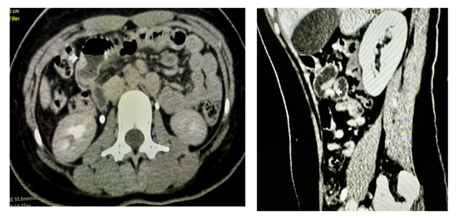
Figure 1: Transverse plane CECT; Figure 2: Sagittal plane CECT.
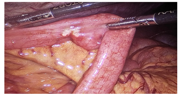
Figure 3: NET of ileum visualised during Diagnostic Laparoscopy.
In view of recurrent intestinal obstruction and imaging findings suggestive of a focal obstructing lesion, the patient underwent diagnostic laparoscopy. Intraoperatively, a localized lesion involving the mid ileum with associated narrowing of the bowel lumen was identified. Segmental resection of approximately 6.5 cm of ileum with primary end-to-end anastomosis was performed.
Gross pathological examination revealed a well-circumscribed submucosal tumour measuring 1 cm in greatest dimension. Microscopic examination demonstrated a well-differentiated neuroendocrine tumour composed of uniform round-to-polygonal cells arranged in nests and trabeculae with characteristic finely stippled (“salt-and-pepper”) chromatin. Mitotic activity was inconspicuous (<2 mitoses per 2 mm²). The tumour infiltrated the submucosa, muscularis propria, and serosa. There was no evidence of lymphovascular invasion or perineural invasion. Surgical resection margins were free of tumour. The pathological stage was pT3N0.
Immunohistochemical analysis showed diffuse positivity for chromogranin A and synaptophysin, confirming neuroendocrine differentiation. The Ki-67 proliferation index was approximately 1%, consistent with a WHO Grade 1 well-differentiated neuroendocrine tumour.
The postoperative course was uneventful, and the patient was discharged in stable condition and has been on regular surgical and oncological follow-up.
DISCUSSION:
Neuroendocrine tumours of the ileum represent the most common primary malignancy of the small intestine and account for nearly 70% of gastrointestinal neuroendocrine tumours. They originate from enterochromaffin cells of the intestinal mucosa and are characterized by slow growth and delayed clinical presentation. Despite their relatively indolent nature, they possess significant metastatic potential, with regional lymph node metastasis identified in more than 50% of patients at diagnosis. [1-3]
The clinical manifestations of ileal NETs are often nonspecific, leading to delayed recognition. Patients commonly present with intermittent abdominal pain, bloating, nausea, weight loss, occult gastrointestinal bleeding, or recurrent episodes of partial intestinal obstruction. [4,5] The obstructive symptoms are frequently disproportionate to tumour size because of the intense desmoplastic reaction induced by tumour-secreted growth factors. This mesenteric fibrosis results in bowel tethering, kinking, and luminal narrowing, producing recurrent subacute obstruction. [6,7]
Cross-sectional imaging plays a pivotal role in diagnosis. Contrast-enhanced CT may demonstrate a hyperenhancing bowel lesion, mesenteric fibrosis, calcification, bowel wall thickening, or features of intestinal obstruction. CT enterography and Ga-68 DOTATATE PET/CT provide superior sensitivity for detecting small primary lesions and occult metastatic disease. [8-10]
Definitive diagnosis relies on histopathological examination and immunohistochemistry. Typical neuroendocrine markers include chromogranin A and synaptophysin, while tumour grading is determined by mitotic activity and the Ki-67 proliferation index according to the WHO classification. Histological grade is one of the most important predictors of prognosis. [11,12]
Surgical resection remains the cornerstone of treatment for localized disease. Complete resection of the primary tumour with associated mesentery and regional lymph nodes offers the best chance of long-term disease control. In patients presenting with bowel obstruction, surgical exploration is often necessary because imaging may underestimate the extent of mesenteric fibrosis and bowel involvement. [13,14]
The present case is noteworthy because the patient was only 27 years old, which is considerably younger than the typical age of presentation, usually occurring during the sixth or seventh decade of life. Furthermore, the tumour measured only 1 cm yet produced recurrent episodes of intestinal obstruction, highlighting the fact that even small ileal NETs can cause significant morbidity through associated fibrotic changes. [15].
CONCLUSION:
Ileal neuroendocrine tumours are uncommon but clinically significant causes of recurrent small bowel obstruction. Their diagnosis is often delayed because symptoms are nonspecific and imaging findings may be subtle. Recurrent or unexplained episodes of intestinal obstruction, particularly in the absence of previous abdominal surgery or other obvious causes, should raise suspicion for an underlying small bowel neoplasm including ileal neuroendocrine tumour. [6,7,13]
Careful radiological evaluation, timely surgical intervention, and histopathological examination are essential for establishing the diagnosis. This case highlights that even small, well-differentiated ileal NETs can present with recurrent obstruction and should be considered in the differential diagnosis of young patients with repeated episodes of subacute intestinal obstruction. [11,13,14].
REFERENCES:
1. Modlin IM, Oberg K, Chung DC, Jensen RT, de Herder WW, Thakker RV, et al. Gastroenteropancreatic neuroendocrine tumours. Lancet Oncol. 2008;9(1):61–72.
2. Yao JC, Hassan M, Phan A, Dagohoy C, Leary C, Mares JE, et al. One hundred years after “carcinoid”: epidemiology of and prognostic factors for neuroendocrine tumours in 35,825 cases in the United States. J Clin Oncol. 2008;26(18):3063–72.
3. Dasari A, Shen C, Halperin D, Zhao B, Zhou S, Xu Y, et al. Trends in the incidence, prevalence and survival outcomes in patients with neuroendocrine tumours in the United States. JAMA Oncol. 2017;3(10):1335–42.
4. Hallet J, Law CHL, Cukier M, Saskin R, Liu N, Singh S. Exploring the rising incidence of neuroendocrine tumours. J Gastrointest Surg. 2015;19(5):824–31.
5. Niederle MB, Hackl M, Kaserer K, Niederle B. Gastroenteropancreatic neuroendocrine tumours: the current incidence and staging based on the WHO and ENETS criteria. Endocr Relat Cancer. 2010;17(4):909–18.
6. Makridis C, Oberg K, Juhlin C, Rastad J, Johansson H, Lörelius LE, et al. Surgical treatment of mid-gut carcinoid tumours. World J Surg. 1990;14(3):377–83.
7. Anthony LB, Strosberg JR, Klimstra DS, Maples WJ, O’Dorisio TM, Warner RR. The NANETS consensus guidelines for diagnosis and management of gastrointestinal neuroendocrine tumours. Pancreas. 2010;39(6):767–74.
8. Pape UF, Perren A, Niederle B, Gross D, Gress T, Costa F, et al. ENETS consensus guidelines for the management of patients with neuroendocrine neoplasms of the jejunum and ileum. Neuroendocrinology. 2012;95(2):135–56.
9. Sadowski SM, Neychev V, Millo C, Shih J, Nilubol N, Herscovitch P, et al. Prospective study of Ga-68 DOTATATE PET/CT in gastroenteropancreatic neuroendocrine tumours. J Clin Oncol. 2016;34(6):588–96.
10. Hope TA, Bergsland EK, Bozkurt MF, Graham M, Heaney AP, Herrmann K, et al. Appropriate use criteria for somatostatin receptor PET imaging in neuroendocrine tumours. J Nucl Med. 2018;59(1):66–74.
11. WHO Classification of Tumours Editorial Board. Digestive System Tumours. 5th ed. Lyon: IARC Press; 2019.
12. Rindi G, Klimstra DS, Abedi-Ardekani B, Asa SL, Bosman FT, Brambilla E, et al. A common classification framework for neuroendocrine neoplasms. Mod Pathol. 2018;31(12):1770–86.
13. Howe JR, Cardona K, Fraker DL, Kebebew E, Untch BR, Wang YZ, et al. The North American Neuroendocrine Tumor Society consensus guideline for management of midgut neuroendocrine tumours. Pancreas. 2017;46(6):707–14.
14. Landry CS, Brock G, Scoggins CR, McMasters KM, Martin RCG. A proposed staging system for small bowel neuroendocrine tumours. J Am Coll Surg. 2008;207(6):874–81.
15. Moertel CG. Karnofsky Memorial Lecture. An odyssey in the land of small tumours. J Clin Oncol. 1987;5(10):1502–22.