Anesthetic Management of Bilateral Inguinal Hernia Repair and Hydrocoele Excision in A Patient with A Permanent Pacemaker - A Case Report.
- Geetha J , Professor, Department of Anaesthesia, Dhanalakshmi Srinivasan Medical College and Hospital, Siruvachur, Perambalur, Tamil Nadu, India.
- Jaseeem Ahamed M , Postgraduate of Anaesthesiology, Dhanalakshmi Srinivasan medical college, Siruvachur, Perambalur, Tamil Nadu, India.
- Reegana Doulath , Postgraduate of Anaesthesiology, Dhanalakshmi Srinivasan medical college, Siruvachur, Perambalur, Tamil Nadu, India.
Article Information:
Abstract:
Background: The perioperative management of surgical patients with cardiac rhythm management devices, such as permanent pacemakers, presents unique challenges to the anesthesiologist. Electromagnetic interference (EMI) generated by surgical electrocautery poses a critical risk of device malfunction, lead failure, or inappropriate mode resetting. This case report describes the successful anesthetic management of a 67-year- old diabetic and hypertensive male with an indwelling permanent dual-chamber pacemaker (in VVI demand pacing mode) scheduled for bilateral inguinal hernia repair and right hydrocoele excision. To avoid the hemodynamic variability associated with general anesthesia or spinal block, regional anesthesia using a titrated epidural technique was selected. Preoperatively, the pacemaker was reprogrammed to an asynchronous VOO mode, and strict bipolar diathermy constraints were implemented. Stable intraoperative hemodynamics were sustained without significant ST-segment or heart rate fluctuations. This report emphasizes the clinical efficacy of structured preoperative pacing device optimization combined with segmental epidural anesthesia to achieve optimal motor block, minimize hemodynamic shifts, and support early postoperative head-up position in. Abbreviations EMI- Electromagnetic Interfernce, VII - Synchronous mode, VOO- Asynchronous mode, CHB- Complete Heart block, METS- Metabolic equivalents, CIED- Cardiac Implantable Electronic Devices.
Keywords:
Article :
INTRODUCTION:
Bilateral inguinal hernia is a chronic structural derangement of the abdominal wall that poses a significant risk of morbidity if left untreated. Over time, progressive enlargement can cause severe discomfort, mechanical impairment during posture changes, and severe complications such as incarceration, strangulation, or bowel obstruction. Surgical repair remains the definitive treatment strategy. However, when presenting in patients with advanced age and significant cardiovascular comorbidities, the anesthetic risks escalate considerably.
CASE HISTORY:
A 67-year-old male presented to the surgical outpatient department presenting with symptomatic bilateral inguinal swelling accompanied by a right- sided scrotal mass, subsequently diagnosed as bilateral inguinal hernia with an associated right hydrocoele.
The patient's medical history was significant for type 2 diabetes mellitus, which was managed via oral hypoglycemic agents, and systemic hypertension, controlled on Tablet Amlodipine for the past six years. Notably, the patient had a profound cardiac history: he underwent permanent cardiac pacemaker insertion 19 years prior (in 2007) following episodes of palpitations and giddiness. At that time, he was diagnosed with atrial flutter that progressed to complete heart block (CHB).
Following the diagnosis of CHB, the patient was stabilized with a permanent pacemaker configured to a ventricular pacing and sensing demand mode (VVI mode). The pacing system underwent a scheduled lead replacement and battery change in 2018. The patient reported a stable functional capacity of greater than 4 Metabolic Equivalents (METs) without recent episodes of syncope, chest pain, or worsening dyspnea.
Clinical Features
On physical examination, the patient was a well- built male with a Body Mass Index (BMI) of 22.2 kg/ m². Visual inspection revealed a distinct, well-healed surgical scar and a visible pacemaker pulse generator device implanted in the left infraclavicular region.
Clinical auscultation and examination revealed no detectable abnormalities within the respiratory or cardiovascular systems. The USG confirmed the presence of a bilateral inguinal hernia containing omentum as the primary herniated content. Additionally, the scan confirmed a right-sided hydrocoele filled with clear fluid, without any internal septations or signs of acute infection.
Investigations
Table 1:
|
Investigation / Parameter |
Value / Finding |
|
Hb (Hemoglobin) |
14.6gm% |
|
Platelets |
2.0 lakhs |
|
Urea |
20mg/dl |
|
Creatinine |
1.1 |
|
Blood Sugar |
(R) 71 mg/dL |
|
Blood Group |
O -ve |
|
PT / INR |
18.8 / 1.1 |
|
APTT |
92.6 sec |
|
BT (Bleeding Time) |
2' 42" |
|
CT (Clotting Time) |
5' |
|
Serology |
NR (Non-Reactive) |
|
ECG |
Pacemaker generated rhythm |
|
CXR (Chest X-ray) |
Pacemaker (L) side in-situ |
|
TFT (Thyroid Function Test) |
T3 -> 0.8, T4 -> 5.3, TSH -> 4.2 |
Table 2
|
Echocardiogram Parameter |
Measured Value |
|
Aortic Root (ED) |
2.2 cm |
|
Left Atrium (ES) |
3.2 cm |
|
LVEF |
55% |
|
FS |
28% |
Table 3
|
Valve |
Velocity (m/sec) |
Gradient (mmHg) |
Regurgitation |
|
Mitral Valve |
E-1.02 A-0.67 |
- |
Moderate |
|
Aortic Valve |
0.93 |
- |
Nil |
|
Pulmonary Valve |
0.98 |
- |
Nil |
|
Tricuspid Valve |
2.62 |
RVSP: 27 mmHg |
Trivial |
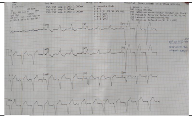
Figure 1
Preoperative Preparation
Given the patient's dependence on a permanent pacemaker and the high likelihood of electrocautery use during bilateral hernia repair, an expert cardiology consultation was secured. To mitigate the risk of electromagnetic interference (EMI)-which could mistake electrocautery signals for intrinsic cardiac activity and inappropriately suppress pacing-the pacemaker mode was converted from its baseline VVI demand mode to an asynchronous ventricular pacing mode (VOO mode) prior to the induction of anesthesia.
The surgical team was strictly instructed to utilize only bipolar diathermy instead of monopolar electrocautery, ensuring the electrical current loop remained confined locally between the instrument tips. On the morning of surgery, Fasting Blood Sugar (FBS), urine ketone levels, and serum potassium concentrations was ordered.
Emergency external defibrillator pads and immediate ionotropic supports (including adrenaline, noradrenaline, and ephedrine) were pre-arranged.
Upon shifting the patient into the surgical holding area on a trolley, standard monitoring was established. Intravenous access was secured with an 18-gauge Venflon catheter, and a maintenance infusion of normal saline was initiated. Premedication was administered via slow intravenous injection of 2 mg of midazolam to alleviate pre-operative anxiety. Comprehensive physiologic monitoring was instituted, consisting of a 5-lead electrocardiogram (ECG) with continuous ST- segment and QT-interval monitoring, Non-Invasive Blood Pressure (NIBP), and pulse oximetry (SpO2).
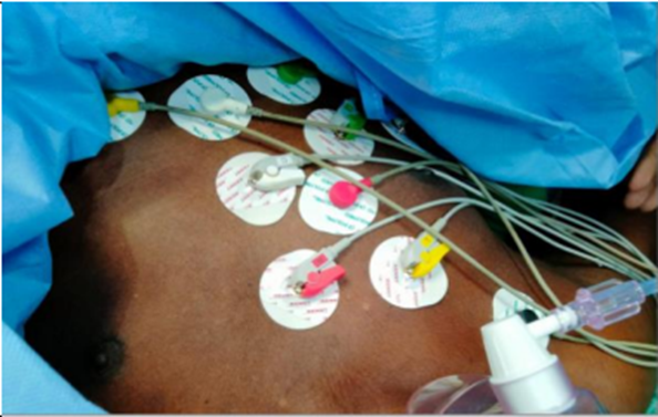
Figure 2
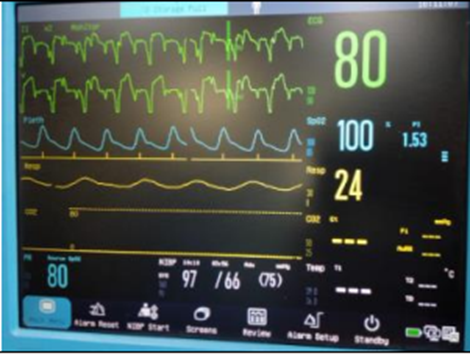
Figure 3
Conduct of Anesthesia
The anesthetic plan was formulated to prioritize regional anesthesia over general anesthesia or standard subarachnoid block. A standalone epidural technique was planned to avoid sudden, profound sympathetic blockades that accompany high spinal anesthesia, which could compromise coronary perfusion in a patient with CAD and Grade 2 LVDD.
Under strict aseptic precautions, with the patient in the sitting posture, an 18G Tuohy epidural needle was inserted into the L3-L4 intervertebral space. The epidural space was identified successfully utilizing the loss-of-resistance technique to saline. An epidural cannula was threaded and fixed at the 15 cm mark on the skin, and an integrated filter was attached.
A localized anesthetic solution was prepared and administered through the catheter, comprising 15 ml of 0.5% Bupivacaine combined with 5 ml of 2% Lignocaine with Adrenaline (1:200,000 dilution). The block was allowed to establish progressively, achieving a bilateral sensory level up to the T10 dermatome, providing excellent abdominal and inguinal analgesia.
To maintain sensory blockade during the bilateral repairs and scrotal dissection, a top-up dose of 7 ml of 0.125% Bupivacaine was administered intraoperatively. Throughout the surgical procedure, the patient's hemodynamic profile remained stable. The ST-segment trended flat, and the heart rate was stably maintained at 70 beats per minute via the asynchronous pacemaker. No episodes of hypotension or significant drops in blood pressure occurred, eliminating the need for vasopressor intervention. Following the completion of surgery, a postoperative ECG was recorded, revealing no acute ST-T wave dynamic changes. The post-operative blood glucose concentration was checked and noted to be stable at 93 mg/dl.
DISCUSSION:
Complete Heart Block (CHB)
Complete atrioventricular block is a high‑grade conduction disturbance associated with syncope, cardiogenic shock, ventricular arrhythmias and increased in‑hospital mortality, especially when occurring with acute coronary syndromes.[1] CHB is a common form of AV block in systemic conditions such as hyperthyroidism and often necessitates pacing, though many lower‑grade blocks can be managed medically.[2,3] Because CHB carries a substantial risk of bradycardia and sudden death, permanent pacing is recommended for symptomatic patients and those with risk features such as profound bradycardia or ventricular dysfunction.[4] It is frequently encountered in the geriatric population.[5,6] When CHB is accompanied by atrial fibrillation, as seen in this 66-year-old patient, the loss of the "atrial kick" further compromises cardiac output and hemodynamic stability.[7-11] Managing such patients requires a thorough understanding of their reduced cardiac reserve and the physiological challenges of aging.
Pacemaker
Atrioventricular block is the most frequent indication for permanent pacemaker implantation after cardiac surgery and in general practice.[12] Long‑term, a significant proportion of patients remain pacemaker‑dependent, highlighting the importance of reliable device function during any subsequent non‑cardiac surgery.[13,14]
The presence of a permanent pacemaker (PPM) is the definitive treatment for symptomatic CHB, ensuring a stable heart rate during physiological stress.[15,16,17] Perioperative management of patients with Cardiac Implantable Electronic Devices (CIEDs) is critical, particularly regarding the risk of electromagnetic interference during surgery.[18,19] It is essential to confirm the device's type, its current programming, and its battery status before any surgical intervention.[20,21] For a patient with a pacemaker, the anesthetic goal is to ensure the device continues to sense and pace effectively despite the surgical environment.[22,23]
Preoperative evaluation should clarify indication for pacemaker, dependency, mode, and need for reprogramming.[24] Permanent pacemakers are “the most effective treatment for symptomatic bradycardia, particularly in elderly patients with complete heart block or sinus node dysfunction.
It is advised to change pacemaker to asynchronous mode (e.g., VOO) before surgery to avoid electromagnetic interference.[25] Use bipolar cautery in short bursts with dispersive pad away from the device.[26]
Continuous ECG and, in high‑risk patients, invasive arterial pressure monitoring are recommended; temporary pacing capability should be available.[27] It is crucial to do the ECG monitoring during the surgery because patients with PPM are at risk of undergoing arrhythmia. After surgery, pacemaker should be reprogrammed to baseline mode and checked before ward transfer.
A detailed perioperative CIED review stresses understanding indications and baseline cardiovascular status,
considering device reprogramming or magnet for high‑EMI surgery establishing secondary pacing methods in case of asystole, intermittent, brief use of cautery with pauses, and immediate communication if inhibition/shocks observed.[28]
Inguinal Hernia
Inguinal hernia repair is one of the most common surgical procedures performed, particularly in older males where abdominal wall integrity often declines.[29] While these procedures are often routine, they can be complicated by the presence of a hydrocele, necessitating a combined surgical approach.[30,31] Standard techniques like the Lichtenstein tension-free hernioplasty are preferred for their efficacy, but the surgical plan must be adapted for patients with significant cardiac comorbidities. In high-risk patients, the priority is to minimize surgical stress and maintain stable central hemodynamics. Mesh hernioplasty (including emergency settings) is associated with faster recovery, shorter hospital stay and lower early recurrence compared with tissue repair, at the cost of slightly higher seroma rates.[32] Laparoscopic techniques, including single‑incision TAPP, have shown low complication and recurrence rates with short hospital stays, confirming inguinal hernia surgery as suitable for elderly patients when systemic risk is controlled.[33]
In high‑risk patients where neuraxial block is contraindicated (coagulopathy, severe stenotic valvular disease, hypovolemia, infection, spinal deformity, pre‑existing neurodeficit, uncooperative), general anaesthesia may be the default but carries risks of delayed recovery, ICU admission, ventilation, and higher morbidity.
Thus in patients with moderate risk cardiac illness, who are posted for noncardiac surgeries, the more preferable and safer alternative is regional anesthesia as it reduces the sympathetic stress response which is present while performing general anesthesia. Ultrasound‑guided ilioinguinal/iliohypogastric/genital branch blocks can avoid GA and provide intra‑operative anaesthesia and analgesia.
Anesthesia Considerations in Patients with Pacemakers
The main perioperative concern in pacemaker patients is electromagnetic interference (EMI) from devices such as monopolar electrocautery, which can inhibit pacing or cause malfunction in CIEDs.[34] Guidelines emphasize pre‑operative device identification and follow‑up, assessment of pacing dependence, and planning of magnet use or reprogramming based on procedure site and EMI risk.
Anesthesia for patients with pacemakers focuses on maintaining stable hemodynamics and avoiding triggers that could interfere with the device. Neuraxial techniques, such as the single-shot epidural used in this case, are often advantageous for cardiovascular patients because they avoid the hemodynamic surges associated with general anesthesia and endotracheal intubation. A paced heart cannot compensate for hypotension by tachycardia and hence spinal anesthesia should be cautiously used.
Epidural anesthesia provides a more gradual onset of sympathetic blockade compared to spinal anesthesia, which helps preserve cardiac stability. Ultrasound‑guided ilioinguinal/iliohypogastric ± genitofemoral blocks have been used as sole anesthesia in high‑risk cardiac patients for hernioplasty, avoiding general and neuraxial anesthesia.
Large series and protocols show these blocks can reliably replace spinal anesthesia for open inguinal hernia repair with good pain control and early discharge but may be insufficient as sole technique in some cases and requires ultrasound and expertise.
Low-dose spinal anesthesia with invasive arterial monitoring was used safely in a 75‑year‑old with DDDR pacemaker for bilateral inguinal hernioplasty; pacemaker was switched to VOO mode and brief phenylephrine infusion maintained blood pressure. Combined lumbar epidural + small-dose spinal bupivacaine with fentanyl was used in a 70‑year‑old with severe LV dysfunction undergoing TURP + bilateral hernioplasty with stable course.
Epidural alone provided adequate anesthesia for bilateral hernia repair in a pacemaker patient with sick sinus syndrome and dementia.[35]
In 100 obese patients having hernioplasty, combined spinal‑epidural with low‑dose bupivacaine produced minimal changes in stroke volume, cardiac output, and cardiac index, while spinal alone caused larger fluctuations.
Elderly patients with severe valvular disease or heart failure undergoing inguinal/umbilical hernia repair under lumbar or segmental epidural also showed stable intraoperative BP and HR when low‑concentration local anesthetic was titrated and vasopressors were available.
Segmental or lower lumbar epidural with low‑concentration ropivacaine/bupivacaine and slow incremental dosing is repeatedly associated with better stability than dense/high blocks. In patients with severe MR, dilated cardiomyopathy, or ASA ≥III, carefully titrated epidural allowed acceptable stability when combined with vigilant invasive monitoring and ready vasopressors.
Anesthetic Techniques for Inguinal Hernia Repair
Inguinal hernia repair can be safely performed under general, regional (spinal/epidural), or local anesthesia with or without sedation, with choice guided by patient comorbidity and surgical approach.
Laparoscopic repairs are conducted under general anesthesia, reflecting pneumoperitoneum and Trendelenburg requirements, whereas open mesh repairs can be performed under neuraxial or local techniques, which may avoid airway manipulation and doses of cardiodepressant drugs in high‑risk cardiac patients.[8] In pacemaker‑dependent patients, neuraxial techniques such as a carefully titrated single‑shot epidural for lower abdominal surgery may be better, provided strict hemodynamic monitoring and CIED‑focused precautions against EMI are applied.[36]
Peripheral nerve blocks, such as ilioinguinal/iliohypogastric or transversus abdominis plane (TAP) blocks, offer excellent localized analgesia with minimal systemic effects. However, a single-shot epidural remains an excellent choice for providing the necessary muscle relaxation and comprehensive field block for both hernioplasty and hydrocele excision in a patient with a permanent pacemaker. The final choice of anesthesia must be tailored to the patient's specific cardiac status and the requirements of the surgical procedure.
Summary
The anesthetic management of patients presenting with permanent pacemakers undergoing non-cardiac surgery requires an integrated understanding of electronic device mechanics and cardiovascular physiology. The primary perioperative challenge stems from electromagnetic interference (EMI) generated by surgical electrocautery units. EMI can be misinterpreted by demand pacemakers (such as VVI) as native ventricular depolarization, leading to the inappropriate inhibition of pacing outputs and potentially causing asystole in pacemaker-dependent patients. In this case, converting the pacing configuration from VVI to an asynchronous VOO mode guaranteed a continuous, uninhibited pacing rate of 70 bpm, independent of any external electrical signals. The mandated execution of bipolar diathermy further restricted the path of electrical currents, keeping it remote from the infraclavicular pulse generator and its transvenous leads.
The choice of anesthetic technique plays an equally vital role in optimizing outcomes. While general anesthesia allows controlled ventilation, it can introduce rapid hemodynamic shifts during induction and emergence, compromising myocardial oxygen supply-demand balances in cardiac patients. Standard subarachnoid spinal blockades, though effective for lower abdominal procedures, can induce rapid, dense sympathetic blocks that result in severe hypotension and reflexive tachycardia, which are highly detrimental in the presence of Grade 2 diastolic dysfunction.
In contrast, a titrated epidural anesthetic technique offers a slow, controlled onset of segment-specific sensory and motor blockade. Furthermore, standalone epidural anesthesia provides excellent abdominal wall relaxation and profound postoperative analgesia without the systemic depressant effects of high-dose opioids. This case demonstrates that an epidural block effectively avoids dangerous hemodynamic lability while allowing safe, early head-up positioning in the immediate post- anesthetic period, which improves diaphragmatic fuction and enhances patient comfort.
CONCLUSION:
This case confirms that combining structured preoperative pacemaker reprogramming (to an asynchronous VOO mode) with a carefully titrated standalone regional epidural block represents a safe and highly effective anesthetic approach for patients with complex cardiac pacing devices undergoing major bilateral inguinal surgeries. This management effectively eliminates the risks of electromagnetic device interference, ensures absolute hemodynamic stability, and prevents adverse postoperative cardiovascular events.
REFERENCES:
1. Jiao J, Zhu X, Zhou C, et al. Single-incision laparoscopic transabdominal preperitoneal hernioplasty: 1,054 procedures and experience. Hernia 2023;27(5):1187-94.
2. Merin O, Ilan M, Oren A, Fink D, Deeb M, Bitrán D, et al. Permanent pacemaker implantation following cardiac surgery: indications and long-term follow-up. Pacing Clin Electrophysiol 2009;32(1):29-35.
3. Thomas H, Plummer C, Wright I, et al. Guidelines for the peri-operative management of people with cardiac implantable electronic devices. Anaesthesia 2022;77(9):1020-31.
4. Bordachar P, Zachary W, Ploux S, et al. Pathophysiology, clinical course, and management of congenital complete atrioventricular block. Heart Rhythm 2013;10(5):760-6.
5. Abreu JB, Amorim A, Gama K. Epidural anesthesia: when the benefits outweigh its risks in cardiovascular patients. Reg Anesth Pain Med 2021;70(Suppl 1):A20.
6. Baheti S, Singh V, Singh P. Segmental epidural anaesthesia with ropivacaine for umbilical hernia repair in a patient with severe cardiac dysfunction: a case report. J Clin Diagn Res 2025;19(5):UD03-5.
7. Wattwil M, Sundberg A, Arvill A, et al. Circulatory changes during high thoracic epidural anaesthesia. Acta Anaesthesiol Scand 1985;29(8):849-55.
8. Sundberg A, Wattwil M. Circulatory effects of short-term hypercapnia during high thoracic epidural anaesthesia in elderly patients. Acta Anaesthesiol Scand 1987;31(2):99-104.
9. Dohi S, Tsuchida H, Mayumi T. Baroreflex control of heart rate during cardiac sympathectomy by epidural anesthesia in lightly anesthetized humans. Anesth Analg 1983;62(9):815-20.
10. Deschamps A, Kaufman I, Backman SB, et al. Autonomic nervous system response to epidural analgesia in laboring patients. Anesthesiology 2004;101(1):21-7.
11. Mavarez AC, Ripat C, Suarez M. Pectoralis plane block for pacemaker insertion: a successful primary anesthetic. Front Surg 2019;6:67.
12. Ata F, Khan HA, Choudry H, et al. A systematic review of the clinical characteristics and course of atrioventricular blocks in hyperthyroidism. Ann Med 2024;56(1):2365405.
13. Taha A, David A, Ragnarsson S, et al. Pacemaker implantation after cardiac surgery: a contemporary, nationwide perspective. Heart 2024;111(1):32-8.
14. Finke J, Pasierski M, Dąbrowski E, et al. Permanent pacemaker implantation following mitral valve surgery. State-of-the-art scoping review. Eur J Cardiothorac Surg 2025;67(1):ezaf210.
15. Rastogi S, Goel S, Tempe DK, et al. Anaesthetic management of patients with cardiac pacemakers and defibrillators for noncardiac surgery. Indian J Anaesth 2015;59(9):586-93.
16. Shishir KR, Patil RV, Holyachi R. Anaesthetic management of two patients with pacemaker in-situ posted for Herniorrhaphy. J Clin Anesth Clin Rep 2019;12(3):142-6.
17. Senthilkumar D, Govindan DK, Prasad TK. Anesthetic management of patient with global left ventricular systolic dysfunction posted for transurethral resection of prostate and bilateral hernioplasty. SBV J Basic Clin Appl Health Sci 2022;5(1):10-2.
18. Selvamani, Nayak MR, Bhairavi TG. Anaesthetic management of a patient with permanent pacemaker for emergency bilateral hernioplasty. RJPBCS 2016;7(6):1239-42.
19. Kang SY, Kim HS, Kang H. Referential flow patterns of injectate in epidural and inadvertent subdural anesthesia: exploring the hemodynamic stability of high-level epidural, subdural and combined epidural-subdural blocks in relation to ASA class. J Pain Res 2025;18:4727-41.
20. Davydov NV, Trukhanova IG, Kutyreva Y. Central noninvasive hemodynamics in combined spinal epidural anesthesia. Messenger Anesthesiol Resuscitation 2022;19(4):46-53.
21. Kapitan TA, Pradhana A, Sinardja C. The role of invasive hemodynamic monitoring in patients with severe mitral regurgitation undergoing herniorrhaphy operation. Bioscientia Med 2023;7(18):3862-8.
22. Theodoraki K, Papacharalampous P, Tsaroucha A, et al. The effect of transversus abdominis plane block on acute and chronic pain after inguinal hernia repair: a randomized controlled trial. Int J Surg 2019;63:63-70.
23. Ushnevych Z, Khomyak V, Lerchuk O. Implementation of ERAS principles in treatment of patients with inguinal hernias repaired with Lichtenstein operation. Br J Surg 2024;111(Suppl 4).
24. Giuffrida M, Nita GE, Biolchini F. Old but gold: the surgeon’s affair to manage inguinal hernia. Surg Tech Dev 2023;12(2):87-98.
25. Hariharan U, Sonowal SJ, Kaur M. Pacemaker and geriatric anaesthesia: a special report. Int J Med Rev Case Rep 2018;2(4):119-23.
26. Oktavia E. Cardiac patients for non-cardiac surgery: anesthetic management in patients with permanent pacemaker. Indones J Cardiol 2020;41(1):32-9.
27. Fuster V, Harrington RA, Narula J, et al eds. Diagnosis and Management of Bradycardias. Chap- 89. In: Hurst's the heart. 14th edn. New York: McGraw-Hill Education 2017:2066-8.
28. Galpalli J, Pandey A, Halgaonkar P. Anaesthetic management of a patient with low ejection fraction on CIED posted for hernioplasty. Int J Sci Res (IJSR) 2023;12(12):1135-7.
29. Braun A, Wakefield J, Kukreja P, et al. Peripheral nerve blockade for open inguinal hernia repair in a patient with severe cardiopulmonary disease. Cureus 2024;16(3):e56646.
30. Pandav A. Ultrasound guided ilioinguinal & iliohypogastric nerve blocks combined with genital branch of genitofemoral nerve block for open inguinal hernia repair. J Med Sci Clin Res 2020;8(7):64-8.
31. Ishikawa E, Shibuya M, Yokoyama A, et al. General anesthesia with a transcutaneous pacemaker for a Noonan syndrome patient with advanced atrioventricular block discovered in the remote period after open-heart surgery: a case report. J Dent Anesth Pain Med 2023;23(2):111-6.
32. Bryant H, Roberts P, Diprose P. Perioperative management of patients with cardiac implantable electronic devices. BJA Educ 2016;16(11):388-96.
33. Patterson T, Beck J, Currie P, et al. Meta-analysis of patient-reported outcomes after laparoscopic versus open inguinal hernia repair. Br J Surg 2019;106(7):826-36.
34. Kim M, Kwon C. Perioperative management of patients with cardiac implantable electronic devices. Korean J Anesthesiol 2024;77(4):306-15.
35. Bakhshi R, Patil SS, Kini P, et al. Sick sinus syndrome with Alzheimer's disease for bilateral hernia repair. J Anaesthesiol Clin Pharmacol 2011;27(2):270-2.
36. Ramirez AY, Howes SA, Sanchez K, et al. Anesthesia and EMI: ensuring safe CIED management during surgical procedures. J Cardiothorac Vasc Anesth 2025;39(6):1534-7.
37. Gropper MA, Eriksson LI, Fleisher LA, et al, eds. Perioperative Management of the Patient with Cardiac Disease. Chap- 34. In: Miller's anesthesia. 10th edn. Philadelphia: Elsevier 2025:1086-7.