Paraneoplastic Dermatomyositis Associated with Endometrial Carcinoma with Calcinosis Cutis: A Case Report.
- Pooja Guruprasad Pai , Resident JR2 MD General Medicine Bombay Hospital and Research centre 12, New Marine Lines, Mumbai 400020.
- Gautam Bhansali , MBBS DNB (Internal Medicine) FCPS FACC FACP Consultant Physician & Intensivist Bombay hospital.
- Saurabh Gautam Chhajed , Resident JR3 MD General Medicine, Bombay Hospital and Research centre 12, New Marine Lines, Mumbai 400020.
- Madhu Hingorani Dhanani , MD (Medicine), IDCCM Associate Consultant Faculty DNB Family Medicine.
- Satish Khadilkar , Dean, Bombay Hospital HOD Neurology, Bombay Hospital.
- Jharna Mahajan , Consultant Neurologist, Bombay Hospital.
Article Information:
Abstract:
Background: Dermatomyositis as a Possible Paraneoplastic Manifestation in a Patient with Endometrial Carcinoma: A Case Report. Objectives: To describe the clinical presentation, diagnostic evaluation, treatment response, and malignancy association of dermatomyositis in a patient with a prior history of endometrial carcinoma. Materials and Methods: A 62-year-old female with a history of endometrial carcinoma treated with total abdominal hysterectomy with bilateral salpingo-oophorectomy followed by chemotherapy and radiotherapy in 2021 presented with progressive proximal muscle weakness and characteristic cutaneous manifestations. Clinical examination, laboratory investigations including muscle enzyme assays, electromyography (EMG), nerve conduction studies (NCS), myositis-specific antibody testing, PET-CT imaging, and histopathological evaluation of a posterior thigh swelling were performed. Results: The patient exhibited heliotrope-like facial rash, V-sign/shawl-sign rash, Gottron’s papules, mechanic’s digits, facial skin tightening, and symmetrical proximal muscle weakness. Laboratory evaluation revealed markedly elevated creatine phosphokinase (CPK), aldolase, and lactate dehydrogenase (LDH) levels. EMG and NCS findings were consistent with proximal inflammatory myopathy. Myositis-specific antibody testing demonstrated positivity for Mi-2A and Mi-2B antibodies. PET-CT was undertaken to assess for malignancy recurrence and possible paraneoplastic association. Histopathological examination of the posterior thigh swelling was suggestive of tumor calcinosis/calcinosis cutis. The patient was treated with pulse methylprednisolone followed by oral corticosteroids and mycophenolate mofetil, resulting in significant clinical and biochemical improvement. Conclusion: Dermatomyositis is a rare autoimmune inflammatory myopathy that may occur as a paraneoplastic syndrome, particularly in adults with a history of malignancy. Early recognition of characteristic clinical features, prompt diagnostic evaluation, and timely initiation of immunosuppressive therapy are essential for improved outcomes. This case underscores the importance of ongoing malignancy surveillance and multidisciplinary management in patients presenting with dermatomyositis.
Keywords:
Article :
INTRODUCTION:
Dermatomyositis is an idiopathic inflammatory myopathy characterized by chronic muscle inflammation and pathognomonic cutaneous manifestations. It affects both adults and children and demonstrates a female predominance. Adult-onset dermatomyositis has a well-established association with malignancy and may present as a paraneoplastic syndrome, particularly in association with ovarian, lung, gastrointestinal, breast, and gynecological malignancies.
The disease typically presents with progressive symmetrical proximal muscle weakness accompanied by characteristic dermatological findings such as heliotrope rash, Gottron’s papules, shawl sign, and V-sign rash. Extramuscular involvement may include pulmonary, cardiac, ocular, and gastrointestinal manifestations. Calcinosis cutis is more common in juvenile dermatomyositis but may rarely occur in adults.
The diagnosis is established through a combination of clinical findings, elevated muscle enzymes, electromyographic abnormalities, autoantibody profiling, imaging, and occasionally muscle or skin biopsy. Early diagnosis and prompt immunosuppressive treatment are crucial in preventing long-term morbidity and improving functional outcomes.
We present a case of likely paraneoplastic dermatomyositis in a patient with previously treated endometrial carcinoma, associated with calcinosis cutis and positive myositis-specific antibodies.
CASE PRESENTATION:
A 62-year-old female from Rajasthan, a known case of Endometrial Cancer status post total abdominal hysterectomy with bilateral salpingo-oophorectomy followed by three cycles of chemotherapy and radiotherapy in 2021, presented with erythematous pruritic rash predominantly involving the face, neck, and upper chest for one year, with worsening over the preceding one month. She also developed facial puffiness during this period.
The patient complained of restricted mouth opening and progressive facial skin tightening for one year. Her husband additionally noticed frequent blinking over the past month, associated with dryness of eyes.
She also reported gradually progressive symmetrical proximal muscle weakness involving the upper limbs more than the lower limbs, associated with myalgia, leading to difficulty in raising her arms, dressing, hip flexion, and rising from a squatting position.
Additionally, she noticed a swelling measuring approximately 2 × 5 cm over the left posterior thigh for 4–5 months. Prior histopathological examination performed outside was suggestive of tumor calcinosis.
There was no history of fever, weight loss, dysphagia, arthralgia, hematuria, or Raynaud’s phenomenon.
Examination
Vital parameters were stable:
· Pulse: 82/min
· Blood pressure: 130/80 mmHg
· SpO₂: 99% on room air
Systemic examination was unremarkable.
Neurological Examination
The patient was conscious and oriented. Cranial nerves were intact. Extraocular movements were full and free. No facial asymmetry, dysarthria, or dysphagia was noted.
Motor examination revealed symmetrical proximal muscle weakness:
|
Muscle Group |
Power |
|
Shoulder abduction |
3/5 |
|
Shoulder flexion/extension |
4-/5 |
|
Elbow flexion/extension |
4+/5 |
|
Wrist flexion/extension |
4+/5 |
|
Hip flexion |
3/5 |
|
Hip extension |
4-/5 |
|
Hip abduction/adduction |
4-/5 |
|
Knee flexion/extension |
4+/5 |
|
Ankle flexion/extension |
4+/5 |
· Tone: Normal
· Deep tendon reflexes: 2+ and symmetrical
· Plantars: Bilateral flexor
· Sensory examination: Normal
· Cerebellar signs: Absent
· Meningeal signs: Absent
· Gait: Normal
Cutaneous Findings
Clinical examination revealed:
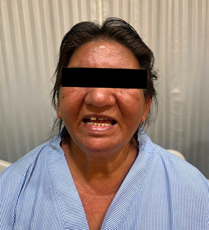
· Heliotrope-like facial rash
· V-sign/shawl-sign rash
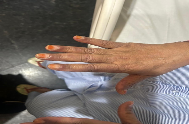
· Gottron's Papules
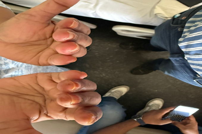
· Mechanic’s digits
· Facial skin tightening
Figure Placeholders
Figure 1. Mechanic’s Digits
Figure 2 Facial sign and v sign
Figure 3. Gottron’s Papules
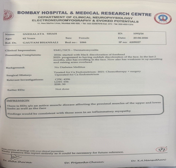
Figure 4. Emg Ncv study
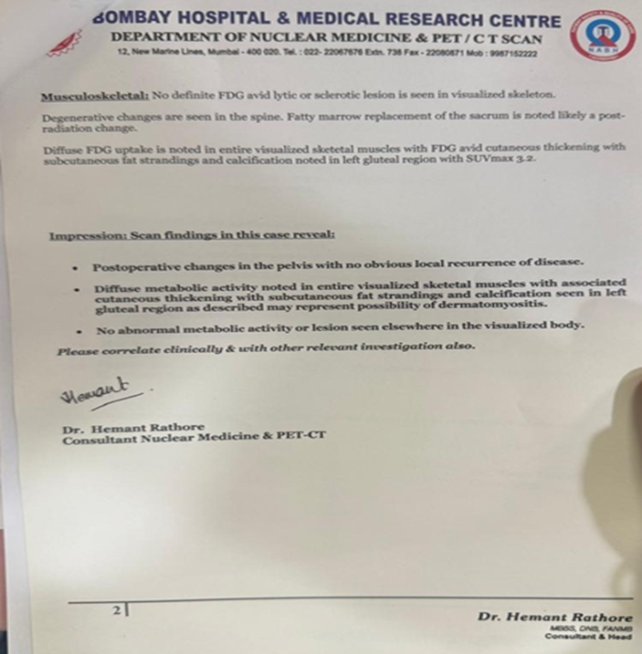
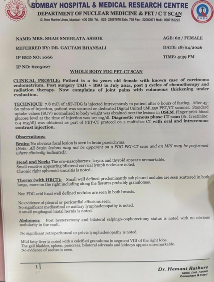
Figure 6. PET-CT Imaging
Investigations
Routine laboratory investigations including complete blood count, liver function tests, and renal function tests were within normal limits.
Muscle Enzymes
|
Investigation |
Value |
|
CPK |
3376 IU/L |
|
CPK-MB |
111.4 IU/L |
|
Aldolase |
25.2 U/L |
|
LDH |
656 IU/L |
|
ESR |
50 mm/hr |
|
CRP |
5.2 mg/L |
Additional investigations:
· Vitamin D: 12.7 ng/mL
· Vitamin B12: >2000 pg/mL
· Serum ACE: 66.14 U/L
Autoimmune Workup
Considering restricted mouth opening, facial skin tightening, glossitis, and dry eyes, autoimmune evaluation was performed to rule out underlying connective tissue disease.
|
Test |
Result |
|
ANA |
Negative |
|
ANA Blot |
Negative |
|
ANCA |
Negative |
|
Myositis Panel |
Mi-2A and Mi-2B Positive |
Imaging
Whole-body PET-CT was performed to evaluate for underlying malignancy recurrence and possible paraneoplastic association.
Histopathology
Posterior thigh swelling biopsy was suggestive of tumor calcinosis/calcinosis cutis.
Electrophysiological Study
EMG-NCS findings were suggestive of proximal inflammatory myopathy.
Management
The patient was evaluated jointly by neurology, dermatology, and ophthalmology teams.
She received:
· Intravenous methylprednisolone pulse therapy for 3 days
· Followed by oral prednisolone (Wysolone) 60 mg/day with gradual tapering
· Mycophenolate mofetil initiated as a steroid-sparing agent
Ophthalmological evaluation including Schirmer’s test revealed mild tear film deficiency bilaterally without superior limbal dryness or congestion.
Supportive management included:
· Lubricating eye drops
· Vitamin D supplementation
· Physiotherapy
· Strict sun protection
· Regular monitoring of CBC, liver function tests, renal function tests, and CPK levels
The patient demonstrated significant improvement in proximal muscle power on repeat neurological examination.
She was discharged on:
· Prednisolone tapering regimen
Mycophenolate mofetil 250 mg twice daily for 2 weeks followed by 500 mg twice daily after laboratory reassessment
DISCUSSION:
Dermatomyositis is associated with malignancy in approximately 15–25% of adult patients. The risk is particularly elevated in elderly patients and in those with dermatomyositis-associated antibodies. Gynecological malignancies including ovarian and endometrial carcinoma have been frequently associated with paraneoplastic dermatomyositis.
The present case is notable for:
• Prior history of endometrial carcinoma
• Characteristic cutaneous manifestations
• Symmetrical proximal muscle weakness
• Elevated muscle enzymes
• Positive Mi-2 antibodies
• Electrophysiological evidence of inflammatory myopathy
• Presence of calcinosis cutis
Calcinosis cutis is uncommon in adult dermatomyositis and is more frequently observed in juvenile disease. Its presence in this patient adds rarity and academic significance to the case.
Mi-2 antibody positivity is generally associated with classic cutaneous manifestations and relatively favorable response to corticosteroid therapy. Early recognition and prompt initiation of immunosuppressive therapy resulted in marked clinical improvement in this patient.
This case underscores the importance of malignancy surveillance in adult-onset dermatomyositis and highlights the need for multidisciplinary management involving neurologists, dermatologists, oncologists, physiotherapists, and ophthalmologists.
CONCLUSION:
This case highlights a rare presentation of likely paraneoplastic dermatomyositis associated with previously treated endometrial carcinoma, complicated by calcinosis cutis. Early diagnosis based on characteristic clinical findings, elevated muscle enzymes, antibody positivity, and electrophysiological evidence allowed timely initiation of immunosuppressive therapy with favorable response. Adult patients presenting with dermatomyositis should undergo thorough malignancy screening and long-term surveillance.
LEARNING POINTS:
· Adult-onset dermatomyositis has a strong association with malignancy.
· Characteristic skin manifestations may precede muscle weakness.
· Calcinosis cutis in adult dermatomyositis is uncommon.
· Myositis-specific antibodies aid diagnosis and prognostication.
· Early immunosuppressive therapy improves outcomes significantly.
· Multidisciplinary care is essential in management.
REFERENCES:
- Lim D, Landon-Cardinal O, Belisle A, Davar S. A case of dermatomyositis with anti-TIF1γ antibodies revealing isolated para-aortic lymphadenopathy metastatic recurrence of endometrial cancer: a case report. SAGE Open Med Case Rep. 2020;8:2050313X20961977. doi:10.1177/2050313X20961977. PMID: 33101685.
- Wada C, Hua CN, Carney ME. Paraneoplastic syndrome in Hawai'i: a case of dermatomyositis associated with endometrial cancer. Hawaii J Med Public Health. 2014;73(4):112–114. PMID: 24765559.
- Jiang W, Yang H, Li S, Tian X, Wang G. Clinical features, treatments and outcomes of calcinosis in adult patients with dermatomyositis: a single cohort study. Rheumatology (Oxford). 2021;60(6):2958–2962. doi:10.1093/rheumatology/keaa618. PMID: 33249499.
- Davuluri S, Chung L, Lood C. Calcinosis in dermatomyositis. Curr Opin Rheumatol. 2024;36(6):453–458. doi:10.1097/BOR.0000000000001036. PMID: 39120537.
- Chandiramani M, Joynson C, Panchal R, Symonds RP, Brown LJ, Morgan B, et al. Dermatomyositis as a paraneoplastic syndrome in carcinosarcoma of uterine origin. Clin Oncol (R Coll Radiol). 2006;18(9):641–648. doi:10.1016/j.clon.2006.07.008. PMID: 17100148.