Lock or Slide? Helical Blade Strategy in Unstable Intertrochanteric Fractures
- Asim Samant , Assistant Professor, Department of Orthopaedics, Kalinga Institute of Medical Sciences, Bhubaneswar, Odisha, India.
- Piyush Sharma , Post-graduate resident, Department of Orthopaedics, Kalinga Institute of Medical Sciences, Bhubaneswar, Odisha, India.
- Basanta Kumar Behera , Professor, Department of Orthopaedics, Kalinga Institute of Medical Sciences, Bhubaneswar, Odisha, India.
Article Information:
Abstract:
Background: Unstable intertrochanteric fractures in elderly osteoporotic patients are associated with high morbidity and mortality. Although cephalomedullary nails with helical blades are widely used, controversy persists regarding the optimal mode of blade fixation—locked (angle- and length-stable) versus dynamic— particularly in terms of fracture collapse, proximal femoral shortening, and functional recovery. Objectives: To compare radiological parameters and functional outcomes between locked helical blade and dynamic helical blade fixation in unstable fragility intertrochanteric fractures. Methods: A prospective observational study was conducted on 60 patients aged ≥55 years with unstable intertrochanteric fractures (AO/OTA 31-A2 and 31-A3). Thirty patients were treated with locked helical blades and thirty with dynamic helical blades using cephalomedullary nails. Radiological outcomes included vertical offset, horizontal offset, neck–shaft angle, and blade back-out. Functional outcome was assessed using the Harris Hip Score (HHS) at 3 and 6 months. Statistical analysis was performed using SPSS v26. Results: The locked helical blade group demonstrated significantly better preservation of vertical offset at 3 months (p = 0.046) and 6 months (p = 0.024). Horizontal offset and neck–shaft angle were better maintained in the locked group, though differences were not statistically significant. Mean HHS at 6 months was higher in the locked blade group, correlating with reduced proximal femoral shortening. Complication rates were comparable, with no significant difference in blade cut-out. Conclusion: Locked helical blade fixation provides superior control of fracture collapse and proximal femoral shortening compared to dynamic blades in unstable intertrochanteric fractures, translating into improved early functional outcomes. Implant selection should be individualized, but locked blades may offer biomechanical advantages in highly unstable osteoporotic fractures.
Keywords:
Article :
INTRODUCTION:
Intertrochanteric fractures constitute a major proportion of hip fractures in the elderly and are predominantly fragility fractures resulting from low-energy trauma in osteoporotic bone. These injuries pose a substantial socioeconomic burden and are associated with high morbidity, loss of independence, and increased mortality [1–4]. The primary objective of treatment is to achieve stable fixation that allows early mobilization, thereby minimizing complications related to prolonged immobilization.
Biomechanically unstable fracture patterns—characterized by posteromedial comminution, lateral wall disruption, or reverse obliquity—are prone to varus collapse and fixation failure. Cephalomedullary nails have emerged as the preferred fixation method for such fractures due to their intramedullary loadsharing design and reduced bending moment compared with extramedullary devices [5–8].
The introduction of helical blades aimed to improve fixation in osteoporotic bone by compacting cancellous bone rather than removing it, thereby enhancing rotational stability [9,10]. However, clinical outcomes with helical blades remain variable, and distinct failure modes such as cut-through and excessive fracture collapse have been described [11–13]. To address uncontrolled sliding and proximal femoral shortening, newer implants offer angle- and length-stable locked helical blade options.
Despite biomechanical rationale, clinical evidence directly comparing locked and dynamic helical blades in unstable intertrochanteric fractures remains limited. Proximal femoral shortening, manifested by changes in vertical and horizontal offset and neck–shaft angle, has been shown to adversely affect gait mechanics and functional outcomes [14–16]. This study aims to compare radiological and functional outcomes between locked and dynamic helical blade fixation in unstable fragility intertrochanteric fractures.
MATERIALS AND METHODS:
Study Design and Setting
A prospective observational cohort study was conducted at a tertiary care teaching hospital between February 2024 and January 2026 after approval from the Institutional Ethics Committee.
The anatomical axis of the femur is defined as a longitudinal line drawn along the femoral shaft, established by joining two mid-diaphyseal points and representing the central axis of the femoral shaft in the coronal plane. The anatomical axis of the femoral neck is defined as a line connecting the centre of rotation of the femoral head to the centre of the femoral neck. The neck–shaft angle is the angle formed between the anatomical axis of the femoral neck and the anatomical axis of the femoral shaft and is a key parameter reflecting proximal femoral alignment. The horizontal offset refers to the perpendicular distance from the centre of the femoral head to the long anatomical axis of the femoral shaft and represents the lateral lever arm of the hip abductors. The vertical offset is defined as the vertical distance between the centre of the femoral head and a horizontal reference line drawn through the lower border of the lesser trochanter or the ischial tuberosity, and it reflects the proximal femoral height and degree of fracture collapse or proximal femoral shortening. (Refer to figure 1)
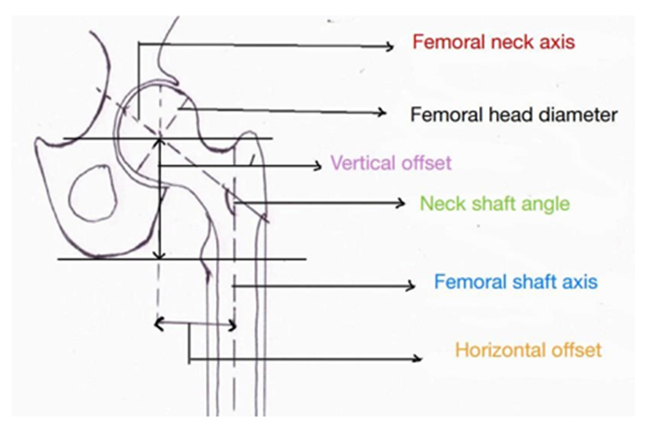
Figure 1. Diagrammatic representation of radiological parameters used to assess proximal femoral geometry, showing the anatomical axes of the femoral shaft and neck, neck–shaft angle, horizontal offset, and vertical offset used for evaluation of proximal femoral shortening
Study Population: -
Sixty consecutive patients aged ≥55 years with unstable intertrochanteric fractures (AO/OTA 31-A2 and 31-A3) resulting from low-energy trauma were included. All patients were ambulatory prior to injury.
Exclusion criteria:
Pathological fractures, polytrauma, previous ipsilateral lower-limb fractures, inflammatory arthritis, neurological disorders affecting gait, and gross postoperative malreduction.
Intervention
Patients were treated with cephalomedullary nails using either:
· Locked helical blade (TFNA)
· Dynamic helical blade (PFNA-2)
Implant choice was based on routine institutional practice. Surgical technique and postoperative rehabilitation protocols were standardized.
Outcome Measures
Radiological assessment included:
· Vertical offset
· Horizontal offset
· Neck–shaft angle
· Blade back-out (%)
Functional outcome was assessed using the Harris Hip Score at 3 and 6 months.
RESULTS:
Demographics
Both groups were comparable in terms of age, sex distribution, fracture pattern, and side of injury. Female predominance was noted, consistent with osteoporotic fragility fractures.
Radiological Outcomes
The locked helical blade group demonstrated significantly better preservation of vertical offset (Refer to figure 2) and neck shaft angle ( Refer to figure 3) at both 3 and 6 months. Progressive reduction in vertical offset was observed in the dynamic blade group, indicating greater fracture collapse. Horizontal offset was better maintained in the locked group, although differences were not statistically significant. (Refer to figure 4).
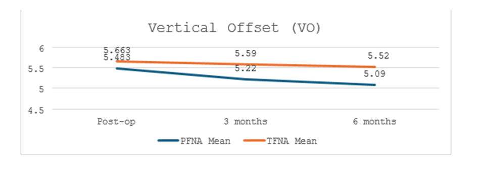
Figure 2. Vertical offset (VO) was assessed postoperatively and at 3- and 6-month follow-up. The dynamic helical blade group demonstrated a gradual reduction in VO over time (5.48 post op to 5.09 at 6 months), indicating progressive collapse. In contrast, the locked helical blade group maintained relatively stable VO values (5.66 post-op to 5.52 at 6 months), with p= 0.046* and 0.024* at 3 and 6 months respectively suggesting superior control of fracture impaction.
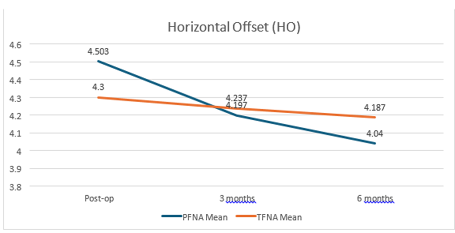
Figure 3: The neck–shaft angle (NSA) was evaluated on standardized anteroposterior radiographs in the immediate postoperative period and at 3- and 6-month follow-up. In the dynamic helical blade group, the mean NSA decreased progressively from 132.21° postoperatively to 129.97° at 3 months and 128.80° at 6 months, indicating gradual varus collapse. In contrast, the locked helical blade group showed minimal change in NSA, with values of 133.25° postoperatively, 132.87° at 3 months (p=0.004*), and 132.47° at 6 months (p=0.001*), reflecting superior maintenance of proximal femoral alignment
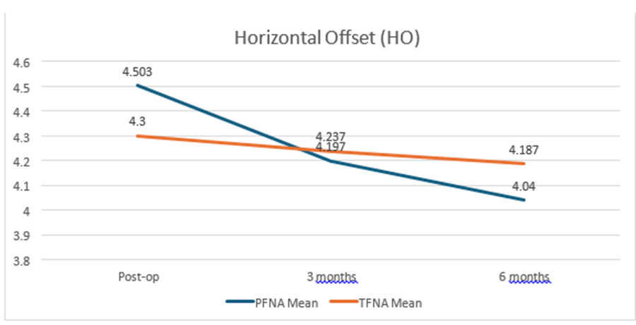
Figure 4:Horizontal offset was assessed on standardized anteroposterior radiographs postoperatively and at 3- and 6-month follow-up. the dynamic helical blade group demonstrated a progressive reduction in mean horizontal offset from 4.50 in the immediate postoperative period to 4.24 at 3 months and 4.04 at 6 months. In contrast, the locked helical blade group showed comparatively minimal reduction in horizontal offset over the same follow-up period (4.30 post-op to 4.19 at 6 months) suggesting comparable reduction between both groups with p=0.435 and 0.556 at 3 and 6 months follow up respectively
Functional Outcomes
Mean HHS improved progressively in both groups. At 6 months, the locked blade group achieved higher mean HHS scores ( Refer Figure 5) , correlating with reduced proximal femoral shortening and better maintenance of hip biomechanics.
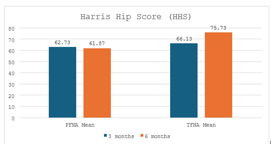
Figure 5. Comparison of mean Harris Hip Score (HHS) between the dynamic helical blade (PFNA-2) and locked helical blade (TFNA) groups at 3- and 6-month follow-up, demonstrating superior functional improvement in the TFNA group at final follow-up
DISCUSSION:
The findings of this study demonstrate that locked helical blade fixation offers superior control of fracture collapse and proximal femoral shortening compared with dynamic blades in unstable intertrochanteric fractures. Preservation of vertical offset is clinically relevant, as excessive shortening compromises abductor lever arm and gait efficiency [14,15].
Dynamic blades permit controlled impaction, which may be beneficial for fracture healing; however, in highly unstable osteoporotic fractures, this can translate into excessive collapse. Locked blades, by providing angular and length stability, better maintain proximal femoral geometry without increasing mechanical failure when optimal reduction and blade positioning are achieved.
Previous biomechanical and clinical studies have reported mixed results when comparing blade designs [17–22]. The present study adds prospective clinical evidence emphasizing radiological parameters that directly influence function rather than focusing solely on union or gross failure.
CONCLUSION:
Locked helical blade fixation in unstable fragility intertrochanteric fractures provides superior control of proximal femoral shortening and better early functional outcomes compared with dynamic helical blades.
While both constructs are effective, locked blades may be preferable in highly unstable osteoporotic fractures where preservation of femoral geometry is critical.
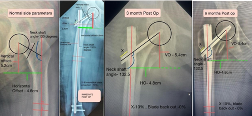
Figure 7. Serial radiographs showing normal side, immediate postoperative, 3-month, and 6-month follow-up images demonstrating maintenance of proximal femoral parameters after locked helical blade fixation.
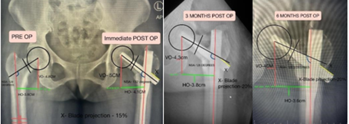
Figure 8 Showing reduction in vertical offset and neck shaft angle leading to varus collapse and blade back out during subsequent follow ups in dynamic helical blade
REFERENCES:
1. Bucholz RW, Heckman JD, Court-Brown CM, Tornetta P. Rockwood and Green’s Fractures in Adults. 9th ed. Philadelphia: Wolters Kluwer; 2020.
2. Bucholz RW, Heckman JD, Court-Brown CM. Rockwood and Green’s
3. Fractures in Adults. 8th ed. Philadelphia: Lippincott Williams & Wilkins; 2015.
4. Court-Brown CM, Caesar B. Epidemiology of adult fractures: A review.
5. Injury. 2006;37(8):691–697.
6. Cooper C, Campion G, Melton LJ. Hip fractures in the elderly: A worldwide projection. Osteoporos Int. 1992;2(6):285–289.
7. Kyle RF, Cabanela ME, Russell TA, et al. Fractures of the proximal part of the femur. J Am Acad Orthop Surg. 1994;2(3):141–153.
8. Hardy DC, Descamps PY, Krallis P, et al. Use of an intramedullary hipscrew compared with a compression hip-screw with a plate for intertrochanteric femoral fractures. Clin Orthop Relat Res. 1998;(348):80–86.
9. Zlowodzki M, Ayieni O, Petrisor BA, Bhandari M. Femoral intramedullary nails versus sliding hip screws for extracapsular hip fractures: A meta-analysis of randomized trials. J Bone Joint Surg Am. 2008;90(10):2154–2165.
10. Bhandari M, Schemitsch E, Jonsson A, Zlowodzki M, Haidukewych GJ.
11. Gamma nails revisited: A systematic review of randomized trials. J Orthop Trauma. 2009;23(6):460–464.
12. Simmermacher RKJ, Bosch AM, Van der Werken C. The Proximal Femoral Nail Antirotation (PFNA): A new device for the treatment of unstable proximal femoral fractures. Injury. 1999;30(5):327–332.
13. Sommer C, Babst R, Muller M, Hanson B. Locking compression plate loosening and plate breakage: A report of four cases. Injury. 2004;35(6):631– 636.
14. Strauss E, Frank J, Lee J, Kummer FJ, Tejwani N. Helical blade versus sliding hip screw for fixation of unstable intertrochanteric hip fractures: A biomechanical evaluation. J Orthop Trauma. 2006;20(7):452–458.
15. Frei HC, Hotz T, Cadosch D, Rudin M, Käch K. Central head perforation, or “cut-through,” caused by the helical blade of the proximal femoral nail antirotation. Clin Orthop Relat Res. 2012;470(8):2278–2287.
16. Yam M, Chui K, Wong K, et al. Mechanical failure of the Proximal Femoral Nail Antirotation device. Injury. 2010;41(12):1231–1236.
17. Madsen JE, Naess L, Aune AK, et al. Dynamic hip screw with trochanteric stabilizing plate in the treatment of unstable proximal femoral fractures. Acta Orthop Scand. 1998;69(5):451–456.
18. Palm H, Jacobsen S, Sonne-Holm S, Gebuhr P. Integrity of the lateral femoral wall in intertrochanteric hip fractures: An important predictor of reoperation. Acta Orthop. 2007;78(5):678–684.
19. Boone C, Carlberg KN, Koueiter DM, et al. Short versus long intramedullary nails for treatment of intertrochanteric femur fractures (OTA 31A1 and A2). J Orthop Trauma. 2014;28(5):e96–e100.
20. Born C, Karich B, Bauer C, et al. Helical blade fixation versus lag screw fixation in osteoporotic bone: A biomechanical comparison. Injury.
21. 2011;42(11):1230–1235.
22. Augat P, Rupprecht M, Weil Y, et al. Bone compaction improves fixation of helical blades in osteoporotic proximal femur fractures. Clin Biomech.
23. 2013;28(8):886–893.
24. Hanke MS, Grobe F, Probst A, et al. Stability of locked versus dynamic helical blade fixation in unstable intertrochanteric fractures. Injury.
25. 2016;47(8):1639–1646.
26. Yoon YC, Lee YK, Kim KC, Ha YC, Koo KH. Cut-through phenomenon after fixation of intertrochanteric fractures using a helical blade. Clin Orthop Surg. 2017;9(4):420–426.
27. Lee SR, Kim ST, Yoon MG, Moon MS, Heo JH. Comparison of helical blade and lag screw fixation for intertrochanteric fractures. Injury.
28. 2015;46(7):1241–1246.
29. Afsari A, Liporace F, Lindvall E, Infante A, Sagi HC. Outcomes of locked versus sliding proximal fixation in cephalomedullary nailing of unstable intertrochanteric fractures. Arch Orthop Trauma Surg. 2018;138(3):349–356.
30. Baumgaertner MR, Curtin SL, Lindskog DM, Keggi JM. The value of the tip-apex distance in predicting failure of fixation of peritrochanteric fractures of the hip. J Bone Joint Surg Am. 1995;77(7):1058–1064.
31. Kuzyk PR, Zdero R, Shah S, Olsen M, Waddell JP, Schemitsch EH. A biomechanical comparison of blade versus screw fixation in a proximal femoral fracture model. J Orthop Trauma. 2012;26(5):e18–e23.
32. Rüedi TP, Buckley RE, Moran CG. AO Principles of Fracture Management. 3rd ed. Stuttgart: Thieme; 2017.