Study of Serum Iron Profile in Patients with Hypothyroidism: A Hospital-Based Case–Control Study.
- Deeksha Swami , Junior Resident, Department of General Medicine, Mahatma Gandhi Medical College and Hospital, Jaipur (Raj.)
- Shrikant Choudhary , Associate Professor, Department of General Medicine, Mahatma Gandhi Medical College and Hospital, Jaipur (Raj.)
- Ram Kishan Jat , Professor, Department of General Medicine, Geetanjali Medical College and Hospital, Jaipur (Raj.)
- Puneet Rijhwani , Professor and Head, Department of General Medicine, Mahatma Gandhi Medical College and Hospital, Jaipur (Raj.).
Article Information:
Abstract:
Background: Iron is an essential cofactor for thyroid peroxidase, and hypothyroidism is closely linked to iron metabolism. Objective: To compare serum iron, ferritin and TIBC in newly diagnosed hypothyroid patients with healthy controls. Methods: A hospital-based case–control study was carried out on 100 subjects (50 cases, 50 controls) aged 18–70 years at Mahatma Gandhi Medical College and Hospital, Jaipur, from February 2024 to July 2025. T3, T4, TSH and ferritin were estimated by CLIA; serum iron by TPTZ; TIBC by Nitroso-PSAP. Data were analysed in SPSS v29 using Student's t-test and Pearson's correlation. Results: Mean serum iron (36.12 ± 10.35 vs 72.57 ± 7.84 µg/dL) and ferritin (24.30 ± 9.67 vs 38.09 ± 6.06 ng/mL) were significantly lower, while TIBC (294.35 ± 62.76 vs 249.61 ± 51.16 µg/dL) and TSH (28.07 ± 5.21 vs 2.98 ± 0.58 mIU/L) were higher in cases (all p < 0.001). Ferritin correlated negatively with TSH (r = −0.53), as did serum iron (r = −0.41). Conclusion: Hypothyroid patients show a clear iron-deficient pattern. Routine iron-profile assessment is recommended in hypothyroidism.
Keywords:
Article :
INTRODUCTION:
The thyroid gland synthesises triiodothyronine (T3) and thyroxine (T4), key regulators of systemic metabolism.¹ Hypothyroidism, characterised biochemically by reduced T3 and T4 with elevated thyroid-stimulating hormone (TSH), affects approximately 5% of adults worldwide and is even more prevalent in Indian cohorts.²,³ Many of its clinical features — fatigue, weight gain, cold intolerance, depression — overlap with those of iron deficiency anaemia, often making clinical differentiation difficult.⁴
Iron metabolism and thyroid hormone synthesis are intimately interlinked. Thyroid peroxidase (TPO), the enzyme that catalyses iodide oxidation and tyrosine iodination to form T3 and T4, is a haem-dependent protein requiring iron as an obligatory cofactor.⁵ Iron deficiency therefore impairs TPO activity, reduces hormone synthesis and elevates TSH compensatorily.⁶ Reciprocally, hypothyroidism itself reduces gastrointestinal iron absorption and erythropoietin-driven iron utilisation, perpetuating iron depletion.⁷
Both conditions are far commoner in women, particularly those of reproductive age, in whom menstrual blood loss and the higher background risk of autoimmune thyroiditis converge.⁸ Several biochemical indices are available to assess iron status: serum iron reflects circulating transferrin-bound iron, ferritin is the principal storage protein and a surrogate of total body iron stores, and TIBC represents the transport potential of transferrin, rising compensatorily during iron depletion.⁹ Despite this well-established bidirectional relationship, iron-profile testing is not routinely performed at the time of hypothyroidism diagnosis. The present study was undertaken to evaluate serum iron, ferritin and TIBC in newly diagnosed hypothyroid patients and to correlate these with thyroid hormones, in comparison with healthy euthyroid controls.
AIMS AND OBJECTIVES
The aim of the study was to evaluate the relationship between iron profile parameters (serum iron, ferritin, TIBC) and the thyroid profile (T3, T4, TSH) in patients with hypothyroidism, in comparison with apparently healthy euthyroid individuals, and to determine correlations between iron and thyroid parameters in the hypothyroid group.
MATERIALS AND METHODS:
This hospital-based case–control study was conducted in the Department of General Medicine, Mahatma Gandhi Medical College and Hospital, Jaipur, between February 2024 and July 2025, after Institutional Ethics Committee approval. One hundred participants (50 newly diagnosed hypothyroid cases and 50 age- and sex-matched healthy controls) aged 18–70 years were enrolled after written informed consent. Patients with other endocrine disorders, chronic kidney disease, chronic infections, recent transfusion, iron supplementation, pregnancy or use of medications affecting thyroid/iron metabolism were excluded.
Approximately 10 mL of fasting venous blood was collected, centrifuged, and serum was separated and analysed within 8 hours. Serum T3, T4, TSH and ferritin were measured by chemiluminescent immunoassay (CLIA); serum iron by the TPTZ method; and TIBC by the Nitroso-PSAP method on a Beckman Coulter AU-400 automated analyser. Data were analysed using SPSS v29. Continuous variables were expressed as mean ± SD; groups were compared with the unpaired Student's t-test, and associations were examined using Pearson's correlation coefficient. A p-value < 0.05 was considered statistically significant.
RESULTS:
The two groups were comparable in age (30.84 ± 10.75 vs 31.84 ± 11.27 years; p > 0.05) and sex distribution (females 70% vs 64%; p > 0.05). The majority of subjects in both groups belonged to the 26–30-year age band, reflecting the demographic in which hypothyroidism most commonly presents (Figures 1 and 2).
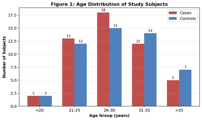
Figure 1. Age distribution of cases and controls.
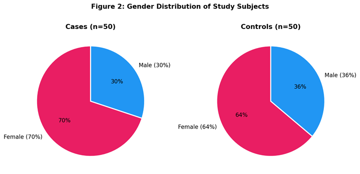
Figure 2. Sex distribution of cases and controls.
Hypothyroid cases showed the expected biochemical derangement: markedly lower T3 and T4 and a sharply elevated TSH, indicating predominantly overt hypothyroidism (Table 1, Figure 1). Iron status was profoundly disturbed: mean serum iron was almost halved, ferritin was reduced by approximately one-third, and TIBC was reciprocally elevated, all changes reaching high statistical significance (Table 1, Figure 2).
Table 1. Comparison of thyroid and iron profile parameters between cases and controls
|
Parameter |
Cases (Mean ± SD) |
Controls (Mean ± SD) |
p-value |
|
T3 (ng/mL) |
0.48 ± 0.20 |
1.42 ± 0.28 |
< 0.001 |
|
T4 (µg/dL) |
2.74 ± 0.68 |
8.07 ± 1.64 |
< 0.001 |
|
TSH (mIU/L) |
28.07 ± 5.21 |
2.98 ± 0.58 |
< 0.001 |
|
Serum iron (µg/dL) |
36.12 ± 10.35 |
72.57 ± 7.84 |
< 0.001 |
|
Ferritin (ng/mL) |
24.30 ± 9.67 |
38.09 ± 6.06 |
< 0.001 |
|
TIBC (µg/dL) |
294.35 ± 62.76 |
249.61 ± 51.16 |
< 0.001 |
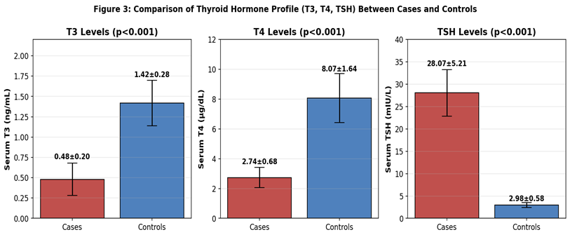
Figure 3. Mean T3, T4 and TSH in hypothyroid cases vs healthy controls.
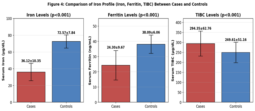
Figure 4. Mean serum iron, ferritin and TIBC in cases vs controls.
Pearson's correlation in the case group (Table 2) revealed that ferritin and serum iron each correlated positively with T3 and T4, and negatively with TSH. The strongest association was the inverse ferritin–TSH correlation (r = −0.53, p < 0.05), illustrated in Figure 5. Serum iron likewise correlated negatively with TSH (r = −0.41, p < 0.05; Figure 6). TIBC correlated negatively with T3 (r = −0.34, p < 0.05) but its relationships with T4 and TSH were weak and non-significant. A consolidated picture of all correlation coefficients is presented in Figure 7.
Table 2. Pearson's correlation between iron profile and thyroid hormones in cases
|
Parameters compared |
Pearson's r |
p-value |
|
Ferritin and T3 |
0.29 |
< 0.05 |
|
Serum iron and T3 |
0.31 |
< 0.05 |
|
TIBC and T3 |
−0.34 |
< 0.05 |
|
Ferritin and T4 |
0.33 |
< 0.05 |
|
Serum iron and T4 |
0.28 |
< 0.05 |
|
TIBC and T4 |
−0.07 |
> 0.05 |
|
Ferritin and TSH |
−0.53 |
< 0.05 |
|
Serum iron and TSH |
−0.41 |
< 0.05 |
|
TIBC and TSH |
0.17 |
> 0.05 |
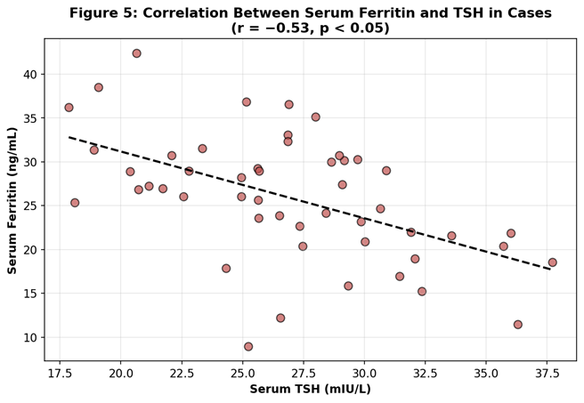
Figure 5. Negative correlation between serum ferritin and TSH in hypothyroid cases (r = −0.53).
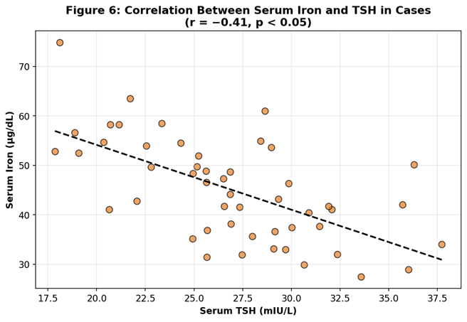
Figure 6. Negative correlation between serum iron and TSH in hypothyroid cases (r = −0.41).
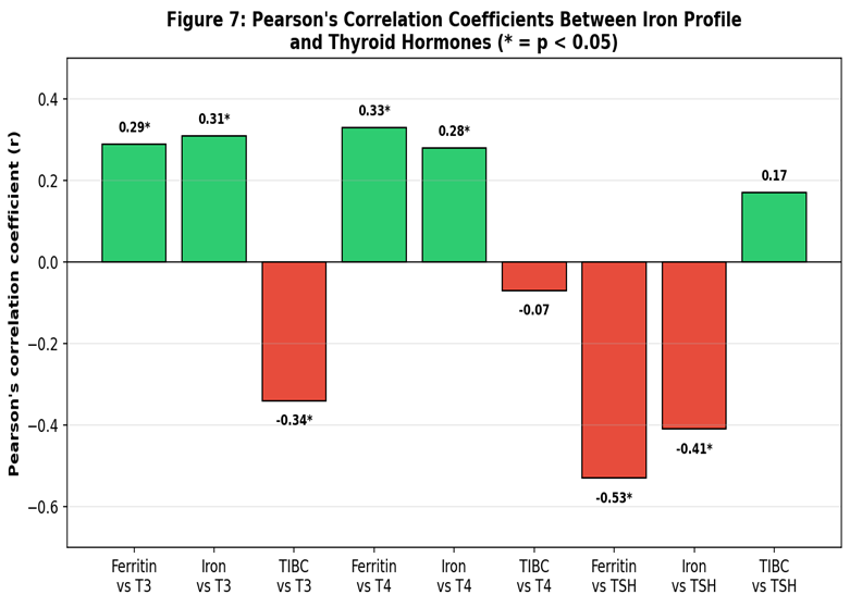
Figure 7. Summary of Pearson's correlation coefficients between iron profile and thyroid hormones (* p < 0.05).
DISCUSSION:
This case–control study demonstrates a clear iron-deficient biochemical signature in newly diagnosed hypothyroid patients — reduced serum iron and ferritin with reciprocally elevated TIBC — together with significant correlations between iron status and thyroid hormones.
The female preponderance (70% of cases) reflects the well-known sex-based predisposition to thyroid dysfunction, particularly autoimmune thyroiditis.⁹ the marked TSH elevation (mean ~28 mIU/L) indicates that most cases had overt rather than subclinical hypothyroidism.
The reduction in serum ferritin and iron, with elevation of TIBC, is consistent with previous reports. Tiwari et al. observed significantly altered ferritin in 143 hypothyroid patients,¹⁰ and Radhakrishnan et al. concluded that iron deficiency aggravates thyroid dysfunction.¹¹ Swapnika et al. demonstrated low ferritin even in subclinical hypothyroidism,¹² and Banday et al. documented a high prevalence of iron-deficiency anaemia in primary hypothyroidism.¹³ Vinayagamoorthi et al.
similarly reported significantly lower iron and ferritin alongside higher TSH in hypothyroid pregnant women.¹⁴ The systematic reviews of Garofalo et al.¹⁵ and Gierach et al.¹⁶ confirm that iron metabolism is consistently disrupted in hypothyroidism. Huang et al., using Mendelian randomisation, established a causal bidirectional link between iron deficiency and hypothyroidism.¹⁷
The mechanistic basis is well characterised. Iron is an obligatory cofactor for TPO; depletion of stored iron (reflected by falling ferritin) impairs iodide oxidation, reduces T3 and T4 synthesis, and elevates TSH compensatorily.⁵,¹⁸ Reciprocally, thyroid hormone deficiency reduces intestinal motility, blunts iron absorption via downregulation of DMT-1 and ferroportin, and decreases erythropoietin output, lowering iron utilisation.⁷ Autoimmune inflammation upregulates hepcidin, sequestering iron in macrophages and enterocytes.¹⁹ This bidirectional loop is summarised in Figure 8.
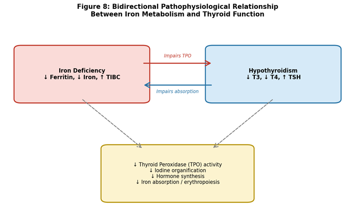
Figure 8. Bidirectional pathophysiological relationship between iron deficiency and hypothyroidism.
The strongest correlation in the present study — ferritin vs TSH (r = −0.53) — is mechanistically intuitive: as iron stores fall, TPO activity declines, hormone synthesis falters and TSH rises. The stronger correlation of T3 (compared with T4) with TIBC may reflect iron-dependent deiodinase activity in peripheral T4-to-T3 conversion.²⁰ Clinically, serum iron and ferritin appear to be more sensitive markers of the iron–thyroid interface than TIBC.
Strengths of this study include the prospective case–control design with well-matched controls and standardised CLIA-based measurements. Limitations include the modest sample size, single-centre recruitment, cross-sectional assessment, and lack of follow-up after thyroxine or iron replacement. Larger multicentric longitudinal studies are required to translate these associations into evidence-based therapeutic protocols, particularly to assess whether concurrent iron correction improves clinical response to levothyroxine.
Clinical Implications
These observations carry several practical implications. First, given that iron deficiency aggravates and is aggravated by hypothyroidism, routine measurement of serum iron, ferritin and TIBC should be considered at the time of initial hypothyroidism diagnosis, especially in women of reproductive age. Second, persistent fatigue or a sluggish response to adequate levothyroxine replacement should trigger evaluation for coexisting iron deficiency rather than escalation of thyroxine alone.
Third, in iron-deficient hypothyroid patients, concurrent oral iron supplementation alongside levothyroxine — administered at separate times to avoid interference with absorption — has been shown to accelerate biochemical and symptomatic recovery.²⁰ Public-health programmes addressing iron deficiency in women of reproductive age may therefore yield collateral benefits in thyroid health, particularly in iodine-replete but iron-depleted populations.
CONCLUSION:
Newly diagnosed hypothyroid patients exhibit significantly reduced serum iron and ferritin with elevated TIBC compared with healthy controls. Iron parameters correlate significantly with thyroid hormones, with ferritin showing the strongest inverse correlation with TSH. Routine iron-profile assessment should be considered in the initial work-up of hypothyroidism, particularly in women of reproductive age, as early identification and correction of coexisting iron deficiency may improve clinical outcomes.
REFERENCES:
1. Carvalho DP, Dupuy C. Thyroid hormone biosynthesis and release. Mol Cell Endocrinol. 2017;458:6–15.
2. Hollowell JG, Staehling NW, Flanders WD, et al. Serum TSH, T4, and thyroid antibodies in the United States population. J Clin Endocrinol Metab. 2002;87(2):489–99.
3. Unnikrishnan AG, Kalra S, Sahay RK, et al. Prevalence of hypothyroidism in adults: an epidemiological study in eight cities of India. Indian J Endocrinol Metab. 2013;17(4):647–52.
4. Vanderpump MPJ. The epidemiology of thyroid disease. Br Med Bull. 2011;99:39–51.
5. Hess SY, Zimmermann MB, Arnold M, Langhans W, Hurrell RF. Iron deficiency anemia reduces thyroid peroxidase activity in rats. J Nutr. 2002;132(7):1951–5.
6. Beard JL, Borel MJ, Derr J. Impaired thyroid function in iron deficiency anemia. Am J Clin Nutr. 1990;52(5):813–9.
7. Dahiya K, Verma M, Dhankhar R, et al. Thyroid profile and iron metabolism: mutual relationship in hypothyroidism. Biomed Res. 2016;27(4):1212–5.
8. Das C, Sahana PK, Sengupta N, et al. Etiology of anemia in primary hypothyroid subjects in a tertiary care center in Eastern India. Indian J Endocrinol Metab. 2012;16(Suppl 2):S361–3.
9. Vanderpump MPJ, Tunbridge WMG. Epidemiology and prevention of clinical and subclinical hypothyroidism. Thyroid. 2002;12(10):839–47.
10. Tiwari R, Saharia GK, Mangaraj M. Evaluation of serum ferritin in hypothyroidism. Endocr Metab Immune Disord Drug Targets. 2021;21(11):2014–9.
11. Radhakrishnan R, Poornima RT, Bawakhan B. Serum ferritin and thyroid profile: a hospital-based study. Asian J Med Sci. 2023;14(3):104–9.
12. Swapnika TG, Rani SS, Dipankar S, Reddy SK. Iron status in subclinical hypothyroidism. Cureus. 2024;16(2):e54221.
13. Banday TH, Bhat SB, Bhat SB, Bashir N, Wani M. Iron deficiency anemia in primary hypothyroidism. Int J Res Med Sci. 2017;5(5):2107–10.
14. Vinayagamoorthi R, Dhiman P, Kollipaka R, Saxena R. Association of hypothyroidism with iron deficiency anaemia in pregnancy. Cureus. 2022;14(10):e30654.
15. Garofalo V, Condorelli RA, Cannarella R, et al. Relationship between iron deficiency and thyroid function: a systematic review and meta-analysis. Nutrients. 2023;15:4790.
16. Gierach M, Rudewicz M, Junik R. Iron and ferritin deficiency in hypothyroidism: a systematic review. Endokrynol Pol. 2024;75(2):109–17.
17. Huang B, Wang L, Liu Y, Zhang H. Bidirectional causal relationship between iron status and thyroid function: a Mendelian randomisation study. Front Endocrinol. 2024;15:1378921.
18. Zimmermann MB, Burgi H, Hurrell RF. Iron deficiency predicts poor maternal thyroid status during pregnancy. J Clin Endocrinol Metab. 2007;92(9):3436–40.
19. Ganz T. Hepcidin and iron regulation, 10 years later. Blood. 2011;117(17):4425–33.
20. Köhrle J. Selenium and the thyroid. Curr Opin Endocrinol Diabetes Obes. 2015;22(5):392–401.