Endoscopic Removal of an Impacted Open Safety Pin in the Postcricoid Region: A Rare and Challenging Foreign Body Case.
- NARALA V HIMAJA REDDY , Postgraduate 3rd year, Department of ENT, Mamata Medical College, Khammam.
- P. SIVA SUBBA RAO , Professor and Head, Department of ENT, Mamata Medical College, Khammam.
- T. NAVEENA , Assistant Professor, Department of ENT, Mamata Medical College, Khammam.
- G. SRAVAN KUMAR , Assistant Professor, DNB Cardiology, Department of Cardiology, Manipal hospitals, Bhubaneshwar.
Article Information:
Abstract:
Background: Foreign body ingestion is a common otorhinolaryngological emergency, but impacted sharp foreign bodies in the adult postcricoid region are uncommon and potentially life-threatening. Open safety pins are especially hazardous because their sharp pointed end may cause mucosal penetration, perforation, deep neck infection, mediastinitis, or migration into adjacent vital structures. We report the case of a 42-year-old female who presented with odynophagia and persistent foreign body sensation in the throat for 20 days, without a clear recollection of ingesting a safety pin. Clinical examination was largely unremarkable, while plain radiographs of the neck revealed an open metallic safety pin at the C5–C6 vertebral level with the pointed end directed laterally. Computed tomography and upper gastrointestinal endoscopy were performed to define the site, orientation, and surrounding tissue involvement. The patient underwent emergency hypopharyngoscopic removal under general anesthesia. The loop of the safety pin was carefully grasped and removed endoscopically without mucosal perforation. Intraoperative purulent discharge was drained, and hemostasis was achieved. The postoperative course was uneventful, with rapid symptomatic improvement and normal follow-up imaging and endoscopic findings. This case highlights the importance of early suspicion, radiological localization, and meticulous endoscopic removal in the management of delayed sharp foreign bodies impacted in the postcricoid region.
Keywords:
Article :
INTRODUCTION:
Foreign body ingestion refers to the presence of any object in the aerodigestive tract where it is not normally meant to be and where it may cause injury by pressure, obstruction, penetration, infection, or migration. Foreign bodies may be endogenous or exogenous, irritant or non-irritant, sharp or blunt, and radiopaque or radiolucent. In otorhinolaryngology practice, commonly encountered foreign bodies include coins, pins, denture wires, batteries, fish bones, chicken bones, mutton bones, plastic toys, beads, and other accidental objects. The common sites of lodgement include the tonsil, base of tongue, vallecula, posterior pharyngeal wall, pyriform fossa, postcricoid region, and esophagus. The cricopharyngeal sphincter is one of the most frequent sites of impaction because it is a natural anatomical narrowing of the upper esophagus (1).
Foreign body ingestion is more common in children; however, adults may also be affected, particularly due to hasty eating, improper mastication, use of dentures, reduced protective airway reflexes, psychiatric illness, alcohol intoxication, or underlying esophageal narrowing such as stricture or carcinoma. Symptoms depend on the site and type of foreign body and may include throat pain, odynophagia, dysphagia, foreign body sensation, drooling of saliva, cough, stridor, dyspnea, wheeze, hemoptysis, voice change, and, rarely, sudden airway compromise. Sharp foreign bodies require particular attention because delayed diagnosis or removal may result in perforation, abscess formation, mediastinitis, vascular injury, or migration into deep neck spaces (2,3).
Open safety pins are rare but hazardous foreign bodies. Their management is technically challenging because the pointed end may injure the mucosa during swallowing, impaction, or extraction. Endoscopic removal is preferred when feasible, but it requires accurate radiological localization, careful assessment of the orientation of the pin, airway protection, and controlled extraction under direct visualization (4,5). The present case describes delayed presentation of an impacted open safety pin in the postcricoid region of an adult female, successfully managed by hypopharyngoscopic removal under general anesthesia.
CASE PRESENTATION:
A 42-year-old female presented to the Department of Otorhinolaryngology, Mamata Medical College, Khammam, with complaints of pain while swallowing and persistent foreign body sensation in the throat for 20 days. The pain was sudden in onset, gradually progressive, intermittent, pricking in character, moderate in intensity, non-radiating, and more pronounced while swallowing solids than liquids. Although there was a history suggestive of ingestion of some foreign body 20 days earlier, the patient did not clearly recall ingestion of a safety pin.
There was no history of choking, gagging, drooling of saliva, respiratory distress, substernal pain, epigastric pain, change in voice, significant dysphagia, decreased taste perception, bleeding from the throat, or symptoms suggestive of gastroesophageal reflux disease. There were no significant ear or nasal complaints. Local throat examination showed missing upper third molars on both right and left sides, while the remaining oral cavity and oropharyngeal examination was unremarkable. Neck examination revealed a midline trachea, preserved laryngeal crepitus, absence of palpable cervical lymph nodes, and no tenderness in the lower neck on either side of the trachea.
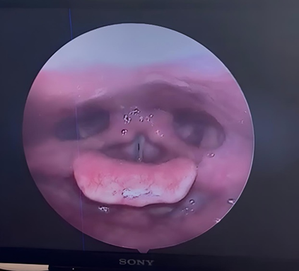
Figure 1: VLS of patient showing Normal vocal cord mobility , no pooling of saliva in pyriform fossa, phonatory gap present.
Videolaryngoscopy showed normal vocal cord mobility, no pooling of saliva in the pyriform fossae, and a phonatory gap. Plain radiographs of the neck in anteroposterior and lateral views revealed a metallic foreign body consistent with an open safety pin at the C5–C6 cervical vertebral level. The base of the safety pin was directed upwards, the hook was directed downwards, and the pointed end was directed laterally. Computed tomography of the neck was performed for precise localization and to assess surrounding soft tissue involvement. Upper gastrointestinal endoscopy was also done to evaluate the foreign body site and mucosal condition.
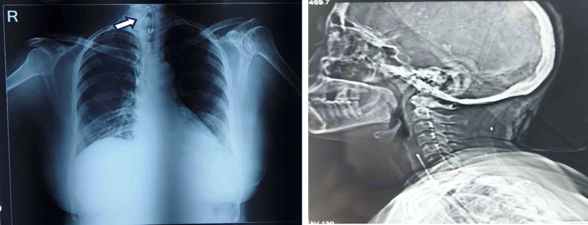
Figure 2: Anteroposterior and lateral radiographs of the neck demonstrating an open metallic safety pin impacted in the postcricoid/cervical esophageal region.
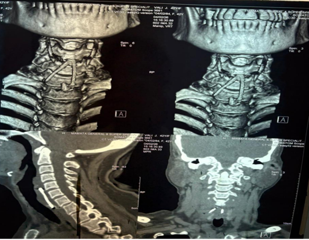
Figure 3: CT neck with 3D reconstruction, sagittal, and coronal views demonstrating an open metallic safety pin impacted at the C5–C6 level in the postcricoid region.
After completing the required investigations, the patient was shifted to the operating theatre for emergency removal under general anesthesia. She was intubated with a 6.5 mm endotracheal tube. After protecting the lips and teeth with guards, a hypopharyngoscope was introduced through the oral cavity. The scope was advanced along the base of tongue, and the uvula and epiglottis were visualized. It was then passed further downwards into the postcricoid region, where the open safety pin was identified under endoscopic visualization.
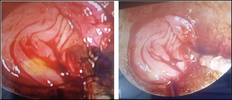
Figure 4: Intraoperative endoscopic images demonstrating inflamed postcricoid mucosa with purulent discharge following impaction of the open safety pin.
The loop of the safety pin was carefully grasped with forceps and extracted safely without causing mucosal perforation. During the procedure, purulent discharge was noted from the 12 o’clock position and was drained. Hemostasis was achieved using lignocaine and adrenaline packs. The hypopharyngoscope was then removed, and the patient was stabilized and extubated. There were no significant immediate postoperative complication.
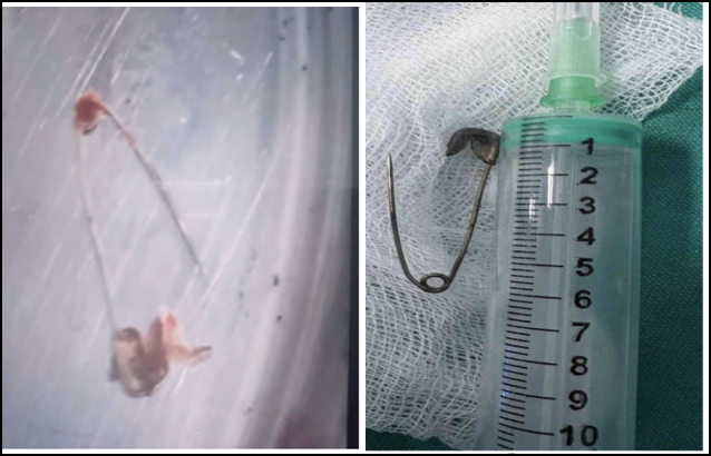
Figure 5: Removed open metallic safety pin with attached soft tissue debris, shown after successful endoscopic extraction from the postcricoid region.
Postoperatively, the patient showed rapid symptomatic improvement. Postoperative radiograph on day 1 showed no retained foreign body. Follow-up upper gastrointestinal endoscopy on postoperative day 10 showed satisfactory healing without significant residual pathology. The patient was monitored postoperatively and discharged after 5 days in stable condition.
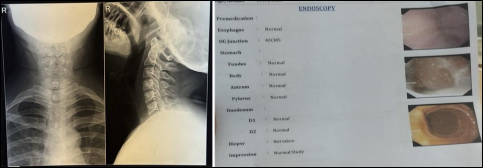
Figure 6: Postoperative anteroposterior and lateral neck radiographs showing no retained foreign body, with follow-up upper GI endoscopy demonstrating normal findings after hypopharyngoscopic removal of the safety pin.
DISCUSSION:
Foreign bodies in the upper aerodigestive tract may present with variable symptoms depending on the age of the patient, type of object, site of impaction, duration since ingestion, and presence of complications. In the esophagus, three anatomical narrowing points are classically described: the cricopharyngeal sphincter, the level where the aortic arch crosses the esophagus, and the lower esophageal sphincter (1). The postcricoid region and cricopharyngeal area are common sites of impaction because of anatomical narrowing and functional sphincteric activity.
Sharp foreign bodies are clinically important because they are more likely to penetrate the mucosa and cause complications than smooth or blunt objects. Potential complications include mucosal ulceration, perforation, retropharyngeal abscess, parapharyngeal abscess, mediastinitis, migration into deep neck spaces, vascular injury, and airway compromise.(2,3). In the present case, the patient had a delayed presentation of 20 days and intraoperative purulent discharge, indicating local inflammatory or infective change. However, timely surgical intervention prevented more serious complications.
Radiological evaluation is essential in cases of suspected sharp foreign body ingestion. Plain anteroposterior and lateral radiographs of the neck are simple, rapid, and useful for detecting radiopaque foreign bodies such as metallic pins. However, radiographs may be insufficient to assess soft tissue extension, perforation, abscess formation, or migration. Computed tomography is therefore valuable, particularly for sharp, penetrating, or delayed foreign bodies, as it helps determine the exact location, orientation, depth of penetration, and relationship to adjacent structures. (2,6). In this patient, radiographs identified the safety pin at C5–C6 level, while computed tomography helped in further assessment and operative planning.
Endoscopic removal is the preferred management for accessible upper aerodigestive tract foreign bodies, especially when the object is sharp, impacted, or symptomatic. Guidelines recommend urgent or emergent endoscopic intervention for sharp-pointed objects in the esophagus because delay increases the risk of perforation and other complications. (7, 8). The technique of removal depends on the type, size, shape, orientation, and site of the object. In the case of an open safety pin, the pointed end poses a major risk during extraction. Therefore, grasping the loop or safer blunt end and removing the pin under direct visualization is essential to avoid additional mucosal trauma.
The present case is notable because an open safety pin was impacted in the postcricoid region for 20 days, yet was removed successfully by hypopharyngoscopy without perforation. The absence of severe symptoms such as drooling, respiratory distress, or overt dysphagia may delay diagnosis in such cases. Therefore, persistent odynophagia or foreign body sensation, even with an unremarkable clinical examination, should prompt imaging. The successful outcome in this case can be attributed to careful preoperative localization, airway protection with endotracheal intubation, controlled endoscopic visualization, grasping of the loop of the safety pin, drainage of purulent discharge, and close postoperative monitoring.
CONCLUSION:
Impacted open safety pins in the postcricoid region are rare but potentially dangerous foreign bodies. Delayed presentation increases the risk of local infection, perforation, migration, deep neck abscess, and mediastinitis. A high index of suspicion is necessary when patients present with persistent odynophagia or foreign body sensation, even when the history is unclear and clinical examination is apparently normal. Plain radiographs are useful for initial diagnosis of metallic foreign bodies, while computed tomography provides accurate localization and assessment of complications. Hypopharyngoscopic removal under general anesthesia is a safe and effective minimally invasive method when performed carefully under direct visualization. Early diagnosis, appropriate imaging, meticulous endoscopic technique, and postoperative monitoring are crucial for successful outcome.
REFERENCES:
1. Baser M, Arslantürk H, Kisli E, Arslan M, Oztürk T, Uygan I, et al. Primary aortoduodenal fistula due to a swallowed sewing needle: a rare cause of gastrointestinal bleeding. Ulus Travma Acil Cerrahi Derg. 2007;13(2):154-7.
2. Nisha V, Prasad KC, Girish I, Mohammadi K. Varied manifestations of sharp penetrating foreign bodies in the aerodigestive tract: our experience. Cureus. 2023;15(5):e39525. doi:10.7759/cureus.39525.
3. ASGE Standards of Practice Committee, Ikenberry SO, Jue TL, Anderson MA, Appalaneni V, Banerjee S, et al. Management of ingested foreign bodies and food impactions. Gastrointest Endosc. 2011;73(6):1085-91.
4. Demiroren K, Kocamaz H, Dogan Y. A case series of ingested open safety pin removal using therapeutic endoscopy in children. Pediatr Gastroenterol Hepatol Nutr. 2019;22(5):441-6.
5. Webb WA, McDaniel L, Jones L. Foreign bodies of the upper gastrointestinal tract: current management. South Med J. 1984;77(9):1083-6.
6. Mathew RP, Sarasamma S, Jose M, Govindan BS, Kurian G, Varkey S, et al. Clinical presentation, diagnosis and management of aerodigestive tract foreign bodies in the adult population: part 1. S Afr J Radiol. 2021;25(1):2022.
7. Birk M, Bauerfeind P, Deprez PH, Häfner M, Hartmann D, Hassan C, et al. Removal of foreign bodies in the upper gastrointestinal tract in adults: European Society of Gastrointestinal Endoscopy clinical guideline. Endoscopy. 2016;48(5):489-96.
8. Mosca S, Manes G, Martino R, Amitrano L, Bottino V, Bove A, et al. Endoscopic management of foreign bodies in the upper gastrointestinal tract: report on a series of 414 adult patients. Endoscopy. 2001;33(8):692-6.