Clinical, Radiological and Microbiological Profile of Patients with Bronchiectasis in a Tertiary Care Center: A Prospective Observational Study
- Jayaprakash B , Professor and HOD, Department of Respiratory Medicine, Sree Mookambika Institute of Medical Sciences, Kulasekharam, India.
- S Praveen Kumar , Junior Resident, Department of Respiratory Medicine, Sree Mookambika Institute of Medical Sciences, Kulasekharam, India.
Article Information:
Abstract:
Background: Bronchiectasis is a chronic respiratory disorder characterized by irreversible bronchial dilatation associated with recurrent infections, persistent airway inflammation, and progressive lung damage. Despite increasing recognition of the disease, data regarding its clinical, radiological, and microbiological characteristics in developing countries remain limited. Aim: To evaluate the clinical presentation, radiological features, pulmonary function abnormalities, and microbiological profile of patients with bronchiectasis attending a tertiary care center. Materials and Methods: This prospective observational study was conducted among 45 patients with HRCT-confirmed bronchiectasis in the Department of Respiratory Medicine of a tertiary care hospital. Detailed clinical history, physical examination, pulmonary function testing, chest radiography, and HRCT thorax were performed in all patients. Sputum samples were subjected to Gram staining, bacterial culture and sensitivity testing, AFB smear, CBNAAT, and mycobacterial culture. Data were analyzed using SPSS software version 25.0. Results: Majority of the patients belonged to the age group of 61–70 years with female predominance. Cough with sputum production was the most common presenting symptom. Obstructive ventilatory defect was observed in 60% of patients. HRCT thorax predominantly showed cystic bronchiectasis with bilateral lower lobe involvement. Pseudomonas aeruginosa was the most commonly isolated organism in sputum culture, accounting for 40% of cases, followed by Klebsiella pneumoniae (26.7%) and Acinetobacter baumannii (11.1%). No bacterial growth was observed in 22.2% patients. All patients were negative for AFB smear and CBNAAT. Conclusion: Bronchiectasis predominantly affects elderly individuals and commonly presents with chronic productive cough and obstructive airway disease. HRCT thorax remains the gold standard for diagnosis, while Pseudomonas aeruginosa is the predominant bacterial isolate associated with the disease.
Keywords:
Article :
INTRODUCTION:
Bronchiectasis is a chronic and progressive respiratory disorder characterized by irreversible dilatation and destruction of the bronchial walls resulting from recurrent infection and persistent inflammation of the airways. The disease is associated with chronic cough, sputum production, recurrent respiratory infections, dyspnea, hemoptysis, and gradual decline in pulmonary function[1]. Although previously considered an orphan disease, bronchiectasis is now increasingly recognized as an important cause of chronic respiratory morbidity worldwide because of improved diagnostic imaging and better clinical awareness[2]. The prevalence of bronchiectasis has shown a rising trend globally, particularly among elderly individuals and females. Studies from Europe, North America, and Asia have demonstrated increasing hospital admissions and healthcare burden related to bronchiectasis[3].
In developing countries such as India, post-infectious causes including pulmonary tuberculosis continue to be major contributors to bronchiectatic lung disease. The high burden of tuberculosis, recurrent lower respiratory tract infections, delayed diagnosis, and inadequate treatment facilities contribute significantly to disease prevalence in the Indian population[4]. Bronchiectasis may result from a wide variety of etiologies including post-tuberculous sequelae, chronic bacterial infections, chronic obstructive pulmonary disease (COPD), allergic bronchopulmonary aspergillosis, immunodeficiency states, connective tissue disorders, and congenital disorders such as primary ciliary dyskinesia and cystic fibrosis. Persistent airway inflammation leads to mucociliary dysfunction, bacterial colonization, and progressive structural damage to the bronchial tree, producing a vicious cycle of infection and inflammation[5].
Clinical manifestations vary depending on the severity and extent of disease. Chronic productive cough is the most common presenting symptom, often associated with mucopurulent sputum production and exertional breathlessness. Physical examination may reveal crackles, wheeze, and digital clubbing. Pulmonary function tests commonly demonstrate obstructive airway disease, although restrictive and mixed patterns may also occur in advanced cases[6]. High Resolution Computed Tomography (HRCT) of the thorax is considered the gold standard for diagnosis and assessment of disease extent, morphology, and lobar involvement. Radiological patterns may include cylindrical, cystic, or varicose bronchiectasis, with lower lobe predominance observed in many patients[7].
Microbiological colonization of the airways plays an important role in disease progression and frequency of exacerbations. Several studies have identified Pseudomonas aeruginosa as the most common pathogenic organism isolated from sputum cultures in bronchiectasis patients and its presence has been associated with increased severity, recurrent exacerbations, poor lung function, and higher mortality[8]. Other commonly isolated organisms include Klebsiella pneumoniae, Haemophilus influenzae, and Acinetobacter species. Knowledge of the microbiological profile is essential for guiding antibiotic therapy and preventing recurrent infections. Previous studies conducted by Dimakou et al., Habesoglu et al., and the EMBARC registry have highlighted the clinical heterogeneity, radiological diversity, and microbiological burden associated with bronchiectasis[6,9]. However, there remains limited data from South India regarding the combined clinical, radiological, and microbiological characteristics of bronchiectasis patients in tertiary care settings. Regional variations in etiology, microbial colonization, and disease severity necessitate local studies to improve understanding and optimize patient management[10]. Therefore, the present study was undertaken to evaluate the clinical presentation, radiological patterns, pulmonary function abnormalities, and sputum microbiological profile among patients with bronchiectasis attending a tertiary care center.
Aim and objectives
Aim of the Study
To evaluate the clinical presentation, radiological patterns, and microbiological profile of patients diagnosed with bronchiectasis in a tertiary care centre.
MATERIALS AND METHODS:
Study Design and Setting
This prospective observational study was conducted in the Department of Respiratory Medicine at a tertiary care teaching hospital over a period of 18 months. The study was undertaken to evaluate the clinical presentation, radiological characteristics, and microbiological profile of patients diagnosed with bronchiectasis attending the outpatient and inpatient departments.
Study Population
The study included adult patients diagnosed with bronchiectasis based on clinical features and radiological confirmation using High Resolution Computed Tomography (HRCT) of the thorax. Both male and female patients above 18 years of age were included in the study.
Inclusion Criteria
· Patients aged ≥18 years.
· Patients with clinically suspected bronchiectasis confirmed by HRCT thorax.
· Patients willing to participate in the study and provide informed written consent.
Exclusion Criteria
· Patients with cystic fibrosis.
· Patients with active pulmonary tuberculosis.
· Patients with bronchogenic carcinoma.
· Patients unable to produce sputum samples for microbiological evaluation.
· Patients unwilling to participate in the study.
Sample Size Calculation
The sample size was calculated using the formula for estimation of proportion in descriptive studies:
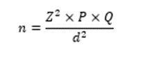
Where:
· n= Required sample size
· Z= Standard normal deviate corresponding to 95% confidence interval = 1.96
· P= Expected prevalence/proportion
· d= Absolute precision
Previous studies on bronchiectasis have demonstrated that Pseudomonas aeruginosa is the most common bacterial isolate in sputum cultures. Stephen Sunny and Mathew Ninan et al. reported a prevalence of Pseudomonas aeruginosa isolation of 43.9% among bronchiectasis patients.
Therefore:
· P=43.9%=0.439
· Z=1.96
· Allowable error d=15%=0.15
Substituting in the formula:
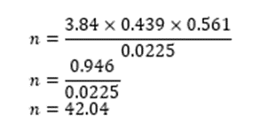
The calculated minimum sample size was approximately 42 patients. Considering possible incomplete data and non-response, the final sample size was rounded to 45 patients.
Sampling Technique
Consecutive sampling method was adopted, and all eligible patients fulfilling the inclusion criteria during the study period were enrolled until the required sample size was achieved.
Data Collection Procedure
After obtaining Institutional Ethics Committee approval and written informed consent, detailed clinical evaluation of all study participants was carried out using a structured proforma.
The following details were recorded:
· Demographic characteristics
· Smoking history
· Past history of tuberculosis, pneumonia, COPD, asthma, or allergy
· Presenting symptoms
· Comorbid illnesses
· Clinical examination findings
Clinical Assessment
Patients were evaluated for symptoms including:
· Cough
· Sputum production
· Dyspnea
· Hemoptysis
· Fever
Dyspnea severity was graded using the Modified Medical Research Council (mMRC) dyspnea scale.
Clinical examination included assessment for:
· Crepitations
· Rhonchi
· Clubbing
Pulmonary Function Testing
Spirometry was performed in all patients using a standardized spirometer according to ATS/ERS guidelines. Pulmonary function patterns were categorized as:
Obstructive Pattern
Restrictive Pattern
Normal Pattern
Severity of obstruction was further classified into mild, moderate, severe, and very severe categories based on predicted FEV1 values.
Radiological Assessment
All patients underwent chest radiography and HRCT thorax.
Chest X-ray Findings
Patients were evaluated for:
· Dilated and thickened bronchi
· Tram-track appearance
· Ring shadows
· Cystic changes
HRCT Thorax Evaluation
HRCT findings were analyzed for:
· Type of bronchiectasis (cylindrical, cystic, varicose, mixed)
· Number of lobes involved
· Distribution of disease
· Unilateral or bilateral involvement
HRCT thorax was considered the gold standard for radiological diagnosis.
Microbiological Assessment
Early morning sputum samples were collected under sterile precautions and sent for:
· Gram staining
· Bacterial culture and sensitivity
· Acid-fast bacilli (AFB) smear
· CBNAAT
· Mycobacterial culture
The predominant bacterial isolates and culture negativity were documented.
Outcome Measures
The primary outcomes assessed were:
· Clinical profile of bronchiectasis patients
· Radiological patterns on HRCT thorax
· Sputum microbiological profile and predominant bacterial isolates
Statistical Analysis
Data were entered in Microsoft Excel and analyzed using Statistical Package for Social Sciences (SPSS) software version 25.0. Categorical variables were expressed as frequency and percentage, while continuous variables were expressed as mean ± standard deviation. Appropriate statistical tests including Chi-square test were used for comparison of categorical variables. A p-value less than 0.05 was considered statistically significant.
Ethical Considerations
The study was initiated after approval from the Institutional Ethics Committee. Written informed consent was obtained from all participants prior to enrollment. Confidentiality and anonymity of patient information were strictly maintained throughout the study period.
RESULTS:
Table 1. Demographic and Clinical Characteristics of Study Participants (n = 45)
|
Variable |
Frequency (n) |
Percentage (%) |
|
Age Group (Years) |
||
|
≤50 years |
6 |
13.3 |
|
51–60 years |
12 |
26.7 |
|
61–70 years |
19 |
42.2 |
|
>70 years |
8 |
17.8 |
|
Gender |
||
|
Male |
20 |
44.4 |
|
Female |
25 |
55.6 |
|
Smoking Status |
||
|
Non-smoker |
28 |
62.2 |
|
Ex-smoker |
13 |
28.9 |
|
Current smoker |
4 |
8.9 |
|
Common Symptoms |
||
|
Cough |
34 |
75.6 |
|
Sputum production |
32 |
71.1 |
|
Dyspnea |
27 |
60.0 |
|
Hemoptysis |
7 |
15.6 |
|
Fever |
5 |
11.1 |
Table 2. Pulmonary Function Test Pattern Among Study Participants (n = 45)
|
Pulmonary Function Pattern |
Frequency (n) |
Percentage (%) |
|
Obstructive pattern |
27 |
60.0 |
|
Restrictive pattern |
7 |
15.6 |
|
Normal pattern |
11 |
24.4 |
Severity of Obstruction (n = 27)
|
Severity |
Frequency (n) |
Percentage (%) |
|
Mild |
4 |
14.8 |
|
Moderate |
7 |
25.9 |
|
Severe |
12 |
44.4 |
|
Very severe |
4 |
14.8 |
Table 3. Radiological Profile of Bronchiectasis Patients (n = 45)
|
Radiological Variable |
Frequency (n) |
Percentage (%) |
|
Chest X-ray Findings |
||
|
Dilated/thickened bronchi |
24 |
53.3 |
|
Tram-track appearance |
10 |
22.2 |
|
Cystic shadows |
11 |
24.4 |
|
HRCT Pattern |
||
|
Cystic bronchiectasis |
21 |
46.7 |
|
Cylindrical bronchiectasis |
17 |
37.8 |
|
Mixed/Varicose pattern |
7 |
15.6 |
|
Distribution |
||
|
Bilateral involvement |
30 |
66.7 |
|
Right-sided involvement |
11 |
24.4 |
|
Left-sided involvement |
4 |
8.9 |
Table 4. Lobar Involvement in HRCT Thorax (n = 45)
|
Lobar Involvement |
Frequency (n) |
Percentage (%) |
|
Lower lobe involvement |
29 |
64.4 |
|
Upper lobe involvement |
26 |
57.8 |
|
Middle lobe involvement |
20 |
44.4 |
Number of Lobes Involved
|
Number of Lobes |
Frequency (n) |
Percentage (%) |
|
Single lobe |
9 |
20.0 |
|
Two lobes |
17 |
37.8 |
|
Three lobes |
12 |
26.7 |
|
≥4 lobes |
7 |
15.6 |
Table 5. Microbiological Profile of Sputum Culture Among Study Participants (n = 45)
|
Sputum Culture Isolate |
Frequency (n) |
Percentage (%) |
|
Pseudomonas aeruginosa |
18 |
40.0 |
|
Klebsiella pneumoniae |
12 |
26.7 |
|
Acinetobacter baumannii |
5 |
11.1 |
|
No growth (KNJM Nil) |
10 |
22.2 |
Additional Microbiological Findings
|
Investigation |
Positive n (%) |
Negative n (%) |
|
Sputum AFB smear |
0 (0%) |
45 (100%) |
|
CBNAAT |
0 (0%) |
45 (100%) |
|
Mycobacterial culture |
0 (0%) |
45 (100%) |
The present study demonstrated that bronchiectasis was more common among elderly female patients. Cough with sputum production was the predominant clinical presentation. Obstructive airway disease was the most frequent spirometric abnormality. HRCT thorax predominantly revealed cystic bronchiectasis with bilateral lower lobe involvement. Pseudomonas aeruginosa was identified as the commonest bacterial isolate in sputum culture, followed by Klebsiella pneumoniae.
DISCUSSION:
Bronchiectasis is a chronic suppurative airway disease associated with recurrent respiratory infections, progressive airway damage, and impaired pulmonary function. The present study evaluated the clinical, radiological, and microbiological profile of 45 patients with HRCT-confirmed bronchiectasis attending a tertiary care center. In the present study, the majority of patients belonged to the age group of 61–70 years with a female predominance. Similar age distribution was reported by Habesoglu et al., who observed increased prevalence of bronchiectasis among elderly individuals due to cumulative airway injury and recurrent infections[6]. Female predominance observed in the present study is also comparable to the findings from the EMBARC registry, which demonstrated a higher prevalence of non-cystic fibrosis bronchiectasis among women[4]. Cough with sputum production was the most common presenting symptom in the current study. These findings are consistent with studies by King et al. and Dimakou et al., who reported chronic productive cough as the hallmark clinical feature of bronchiectasis[2,9]. Dyspnea was observed in 60% of patients in the present study, reflecting progressive airflow limitation and chronic airway inflammation. Hemoptysis was less common and was noted in a smaller proportion of cases, similar to previous Indian studies on bronchiectasis[11].
Pulmonary function testing in the present study predominantly demonstrated an obstructive ventilatory defect in 60% of patients. Comparable observations were made by Lynch et al. and Habesoglu et al., who reported obstructive airway disease as the most frequent spirometric abnormality among bronchiectasis patients[6,7].Chronic airway inflammation, mucus plugging, and bronchial wall destruction contribute significantly to airflow obstruction in these patients. Restrictive and normal pulmonary function patterns observed in a minority of cases may be attributed to localized disease and varying disease severity. Radiologically, cystic bronchiectasis was the most common HRCT pattern identified in the present study, followed by cylindrical bronchiectasis. Bilateral and lower lobe predominance were also commonly observed. Similar HRCT findings were described by Dimakou et al., who reported bilateral multilobar disease with cystic changes in advanced bronchiectasis[9]. HRCT thorax remains the gold standard imaging modality for diagnosis because of its superior ability to detect airway dilatation, bronchial wall thickening, and extent of lobar involvement[7]. Microbiological analysis of sputum cultures in the present study revealed Pseudomonas aeruginosa as the predominant bacterial isolate, followed by Klebsiella pneumoniae and Acinetobacter baumannii. Similar findings were reported by Chalmers et al., who identified Pseudomonas aeruginosa colonization as a major predictor of disease severity, frequent exacerbations, hospitalization, and poor prognosis in bronchiectasis patients[8].
Indian studies conducted by Dhar et al. also demonstrated Pseudomonas species as the commonest respiratory pathogen among patients with non-cystic fibrosis bronchiectasis[4]. The high prevalence of gram-negative organisms in the present study may reflect chronic colonization and repeated antibiotic exposure. No evidence of active tuberculosis was detected in sputum AFB smear, CBNAAT, or mycobacterial culture among the study participants. Similar observations were noted in studies evaluating stable bronchiectasis patients after exclusion of active pulmonary tuberculosis. However, previous pulmonary infections including tuberculosis remain an important etiological factor in the Indian population[4]. Overall, the findings of the present study are consistent with previously published literature and emphasize that bronchiectasis remains an important chronic respiratory disease associated with significant clinical and microbiological burden. Early diagnosis using HRCT thorax, pulmonary function assessment, and microbiological evaluation can facilitate appropriate management and help reduce disease progression and recurrent exacerbations.
Limitations
The study was conducted in a single tertiary care center with a relatively small sample size, which may limit generalization of the findings to the broader population. Etiological evaluation for all causes of bronchiectasis could not be performed uniformly in all patients due to resource limitations. Long-term follow-up to assess exacerbation rates and disease progression was also not included in the study.
CONCLUSION:
Bronchiectasis is a significant chronic respiratory disease predominantly affecting elderly individuals and commonly presenting with chronic cough, sputum production, and obstructive airway disease. HRCT thorax remains the gold standard for diagnosis and assessment of disease extent. The present study demonstrated that cystic bronchiectasis with bilateral lower lobe involvement was the most frequent radiological pattern. Pseudomonas aeruginosa was identified as the predominant bacterial isolate in sputum cultures. Early diagnosis, microbiological evaluation, and appropriate management are essential to reduce recurrent infections, prevent disease progression, and improve patient outcomes.
REFERENCES:
1. Bronchiectasis - an overview | ScienceDirect Topics [Internet]. [cited 2026 May 24]. Available from: https://www.sciencedirect.com/topics/pharmacology-toxicology-and-pharmaceutical-science/bronchiectasis
2. King P. The pathophysiology of bronchiectasis. COPD. 2009;411. doi:10.2147/COPD.S6133
3. Weycker D, Hansen GL, Seifer FD. Prevalence and incidence of noncystic fibrosis bronchiectasis among US adults in 2013. Chron Respir Dis. 2017;14(4):377–84. doi:10.1177/1479972317709649
4. Dhar R, Singh S, Talwar D, Mohan M, Tripathi SK, Swarnakar R, et al. Bronchiectasis in India: results from the European Multicentre Bronchiectasis Audit and Research Collaboration (EMBARC) and Respiratory Research Network of India Registry. The Lancet Global Health. 2019;7(9):e1269–79. doi:10.1016/S2214-109X(19)30327-4
5. Inflammation: a two-edged sword--the model of bronchiectasis. [Internet]. [cited 2026 May 24]. Available from: https://reference.medscape.com/medline/abstract/3533593
6. Habesoglu M, Ugurlu A, Eyuboglu F. Clinical, radiologic, and functional evaluation of 304 patients with bronchiectasis. Ann Thorac Med. 2011;6(3):131. doi:10.4103/1817-1737.82443
7. Lynch DA, Newell J, Hale V, Dyer D, Corkery K, Fox NL, et al. Correlation of CT findings with clinical evaluations in 261 patients with symptomatic bronchiectasis. American Journal of Roentgenology. 1999;173(1):53–8. doi:10.2214/ajr.173.1.10397099
8. Chalmers JD, Goeminne P, Aliberti S, McDonnell MJ, Lonni S, Davidson J, et al. The Bronchiectasis Severity Index. An International Derivation and Validation Study. American Journal of Respiratory and Critical Care Medicine. 2014;189(5):576–85. doi:10.1164/rccm.201309-1575OC
9. Dimakou K, Triantafillidou C, Toumbis M, Tsikritsaki K, Malagari K, Bakakos P. Non CF-bronchiectasis: Aetiologic approach, clinical, radiological, microbiological and functional profile in 277 patients. Respiratory Medicine. 2016;116:1–7. doi:10.1016/j.rmed.2016.05.001
10. Chandrasekaran R, Mac Aogáin M, Chalmers JD, Elborn SJ, Chotirmall SH. Geographic variation in the aetiology, epidemiology and microbiology of bronchiectasis. BMC Pulm Med. 2018;18(1):83. doi:10.1186/s12890-018-0638-0
11. Bronchiectasis - an overview | ScienceDirect Topics [Internet]. [cited 2026 May 24]. Available from: https://www.sciencedirect.com/topics/pharmacology-toxicology-and-pharmaceutical-science/bronchiectasis
12. King P. The pathophysiology of bronchiectasis. COPD. 2009;411. doi:10.2147/COPD.S6133
13. Weycker D, Hansen GL, Seifer FD. Prevalence and incidence of noncystic fibrosis bronchiectasis among US adults in 2013. Chron Respir Dis. 2017;14(4):377–84. doi:10.1177/1479972317709649
14. Dhar R, Singh S, Talwar D, Mohan M, Tripathi SK, Swarnakar R, et al. Bronchiectasis in India: results from the European Multicentre Bronchiectasis Audit and Research Collaboration (EMBARC) and Respiratory Research Network of India Registry. The Lancet Global Health. 2019;7(9):e1269–79. doi:10.1016/S2214-109X(19)30327-4
15. Inflammation: a two-edged sword--the model of bronchiectasis. [Internet]. [cited 2026 May 24]. Available from: https://reference.medscape.com/medline/abstract/3533593
16. Habesoglu M, Ugurlu A, Eyuboglu F. Clinical, radiologic, and functional evaluation of 304 patients with bronchiectasis. Ann Thorac Med. 2011;6(3):131. doi:10.4103/1817-1737.82443
17. Lynch DA, Newell J, Hale V, Dyer D, Corkery K, Fox NL, et al. Correlation of CT findings with clinical evaluations in 261 patients with symptomatic bronchiectasis. American Journal of Roentgenology. 1999;173(1):53–8. doi:10.2214/ajr.173.1.10397099
18. Chalmers JD, Goeminne P, Aliberti S, McDonnell MJ, Lonni S, Davidson J, et al. The Bronchiectasis Severity Index. An International Derivation and Validation Study. American Journal of Respiratory and Critical Care Medicine. 2014;189(5):576–85. doi:10.1164/rccm.201309-1575OC
19. Dimakou K, Triantafillidou C, Toumbis M, Tsikritsaki K, Malagari K, Bakakos P. Non CF-bronchiectasis: Aetiologic approach, clinical, radiological, microbiological and functional profile in 277 patients. Respiratory Medicine. 2016;116:1–7. doi:10.1016/j.rmed.2016.05.001
20. Chandrasekaran R, Mac Aogáin M, Chalmers JD, Elborn SJ, Chotirmall SH. Geographic variation in the aetiology, epidemiology and microbiology of bronchiectasis. BMC Pulm Med. 2018;18(1):83. doi:10.1186/s12890-018-0638-0
21. Pasteur MC, Bilton D, Hill AT, on behalf of the British Thoracic Society Non-CF Bronchiectasis Guideline Group. British Thoracic Society guideline for non-CF bronchiectasis. Thorax. 2010;65(7):577–577. doi:10.1136/thx.2010.142778
22. Pasteur MC, Bilton D, Hill AT, on behalf of the British Thoracic Society Non-CF Bronchiectasis Guideline Group. British Thoracic Society guideline for non-CF bronchiectasis. Thorax. 2010;65(7):577–577. doi:10.1136/thx.2010.142778.